Surgical Specialties B - Ophthamology Year 4 Assessment Questions (30 questions in total with added info from me) Flashcards
https://s3.amazonaws.com/classconnection/403/flashcards/11907403/png/ppngjpgpngjpg-1707C7D4E501E512F89.png

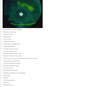
The findings are consistent with wet-age related macular degeneration Intra-vitreal anti-VEGF injections are used as treatment (anti-vascular endothelial growth factor)
What are the symptoms of age related macular degeneration? Which symptom and which sign (on fundoscopy) can indicate the progression from dry to wet age related macular degeneration?
* Initially there is no change in visual acuity, progresses to rapid visual loss secondary to choroidal neovascularisation * Blurred vision * Difficulty with reading and making out faces * Difficulty with seeing at night * As ARMD affects the macula, its effects are on the central vision - central scotoma * Onset of visual distortion (metamorphopsia) can indicate the progression from dry to wet form and requires urgent ophthalmological assessment
What are the deposits that form on the retina / behind the macula in dry ARMD? What is the treatment for dry ARMD?
Drusen spots form on the retina Preventation is the best treatment method - ie no treatment is readily available Antioxidant vitamin and mineral supplement is recommended

What are the risk factors for ARMD?
Smoking Cardiovascular disease Family history Increasing age
What is the first line active treatment of wet ARMD?
1st line - intrravitreal anti-vegF treatment is recommended Ranibizumab - anti-angiogenic antibody - can be given as 1st line for ARMD
What can be offered as an adjunct to anti-VEGF as second-line treatment for late wet ARMD?
Photodynamic therapy can be offered as an adjunct to anti-VEGF for second line treatment of wet-ARMD
https://s3.amazonaws.com/classconnection/403/flashcards/11907403/png/ppngjpg-1707CC11FF86E9C57D7.png

This patient presents with symptoms consistent with thyroid eye disease, along with symptoms to suggest she may have hyperthyroidism. The correct answer is: Thyroid function test and thyroid autoantibodies
How can the inflammatory changes / hypertrophy of certain orbital contents in thyroid eye disease lead to diplopia, proptosis and a dry eye? How can the optic nerve be damaged in this disease?
Thyroid eye disease produces inflammatory changes in the extra-ocular muscles and hypertrophy of the orbital fat, leading to ocular dysmotility and diplopia, proptosis and consequent exposure of the cornea leading to a dry eye. Proptosis and congestion of the orbital contents can be sufficient to stretch the optic nerve and lead to vision loss.
https://s3.amazonaws.com/classconnection/403/flashcards/11907403/png/ppngjpgpngjpgpngjpg-1707CC3A2B24941F457.png

In patients who have tried multiple pharmacological agents to treat the open angle glacuoma, non pharmaoclogical treatments can then be considered Laser trabeculoplasty is usually the next step followed by surgical trabeculectomy for this question, TRABECULECTOMY is the answer
https://s3.amazonaws.com/classconnection/403/flashcards/11907403/png/ppngjpgpngjpg-1707CCEAB8B2ECAF0B6.png

In glaucoma, there is loss of nerve fibres which can be seen as an increase in the cup to disc ratio as axons on the inside of the pale orange halo are lost. This would correspond to a loss of visual field and the formation of scotomas (blind spots), evident on visual field testing. * 1. Glaucoma * 2. Latanoprost (prostoglandin analogue) - increases uveoscleral outflow and timolol (beta blocker) - decreases production of aqueous fluid
https://s3.amazonaws.com/classconnection/403/flashcards/11907403/png/ppngjpgpngjpgpngjpgpngjpg-1707CD2D28B4FF1973A.png

- Cataract extraction 2. Corneal incision, open anterior capsule, phacoemulsification of lens, removal of cortical lens matter, injection of lens implant 3. Where complications arise, these are most commonly related to damage to intra-ocular structures e.g. the lens capsule or iris. Less common complications include an unpredicted refractive error, intra-ocular infection (endophthalmitis) and retinal detachment (which can occur several years after the initial surgery).
Is cataract surgery a complicated procedure?
Cataract surgery is considered to be an extremely safe and effective procedure, with approximately 97% of patients having an uncomplicated operation and recovery.
https://s3.amazonaws.com/classconnection/403/flashcards/11907403/png/ppngjpgpngjpgpngjpg-1707CD6CE18646A54BC.png

Impaired lateral movement of left eye Signifies damaged left abuducens nerve
Giant cell arteritis (GCA) is one of the few true ophthalmic emergencies as it can lead to irreversible and bilateral blindness if left untreated. Jaw/tongue claudication, scalp tenderness, recent weight loss and feeling systemically unwell in a patient over 50 years should warrant investigation for GCA. * What are the visual changes that can occur in GCA? * What tests are carried out?
Visual changes - sudden loss of vision, blurred vision, diplopia, amaurosis fugax Patients should be immediately referred to hospital for blood tests for inflammatory markers, including FBC, CRP and plasma viscosity/ESR.
https://s3.amazonaws.com/classconnection/403/flashcards/11907403/png/ppngjpgpngjpgpngjpg-1707CDC612A242705AF.png

Confirmed GCA is treated with high dose oral steroid, tapering over many months. In cases where there is diagnostic uncertainty, biopsy of the temporal artery may be carried out under local anaesthetic to look for inflammatory changes in the artery wall.
https://s3.amazonaws.com/classconnection/403/flashcards/11907403/png/ppngjpg-1707CDD38275DC6DEF6.png
Dendritic corneal ulcer due to herpetic infection of the cornea Treated with topical acicylovir In ophthalmic herpes simplex infections, there is a risk that topical steroid may lead to enlargement of the ulcer due to localised immunosuppression and worsening infection. This is not a risk with use of oral corticosteroids however.
https://s3.amazonaws.com/classconnection/403/flashcards/11907403/png/ppngjpgpngjpg-1707CDEDB3226A9D957.png

RETINAL DETACHMENT Sudden onset of flashes and floaters and a “curtain-like” shadow or veil across the vision are highly suggestive of retinal detachment which require urgent ophthalmology assessment and potential surgery in order to preserve vision.
Is retinal detachment linked to myopia or hypermetropia?
Retinal detachment is linked to myopia
https://s3.amazonaws.com/classconnection/403/flashcards/11907403/png/ppngjpgpngjpgpngjpg-1707CF22310215032B0.png

Corneal transplant Keratoconus - disorder of the eye which results in progressive thinning of the cornea. - can get bulging of the anterior chamber Corneal scarring following infection or trauma Astigmatism - cornea fails to have the same curvature Graft rejection

https://s3.amazonaws.com/classconnection/403/flashcards/11907403/png/ppngjpgpngjpgpngjpg-1707CF5FD010C4808EB.png

* Branch retinal vein occlusion (BRVO) is occlusion of a tributary to the central retinal vein. It causes the appearance of dilatation in the affected vein with flame shaped, dot and blot haemorrhages. * The patient may present with sudden painless loss of vision or visual field defect The most common sight-threatening complication of a BRVO is macular oedema which reduces central vision. Rarely, neovascularision similar to the process of proliferative diabetic retinopathy can result, where abnormal new vessels may grow in the affected areas of retina.
Why can rubeotic glaucoma occur after BRVO?
When there is abnormal VEGF production (which can occur post-BRVO), these factors can spread into the trabecular meshwork and new blood vessels can grow blocking the drainage of aqueous humour - glaucoma Rubeosis is when there is the formation of abnormal blood vessels
https://s3.amazonaws.com/classconnection/403/flashcards/11907403/png/ppngjpgpngjpgpngjpgpngjpg-1707CF9FACD7572513C.png

Conjunctivitis with a watery discharge and recent coryzal illness suggest viral conjunctivitis which is normally self limiting. Patients should be advised to take precautions to avoid spread to others ie. stay home from school while discharge and redness persist, frequent hand-washing, avoid sharing towels/cosmetics, avoid eye-rubbing. ADVISE COOL COMPRESS / LUBRICANTS
When may you think of a bacterial conjunctivitis? What is the treatment?
Bacterial conjunctivitis usually has Purulent discharge however is also self limiting 1st line is to advise cool compress/lid hugiene/ clean eyes regularly Chloramphenicol is recommended as 1st line antibitoic therapy
A patient with primary open angle glaucoma is started on latanoprost eye drops to reduce her intraocular pressure. What is its main mode of action? What type of drug is latanoprost? Name side effects of this drug class?
Latanoprost is a prostoglandin analogue They Increase aqueous absorption by the uveoscleral outflow Side effects include hyperpigemtnation of the iris/lashes/skin, eyelash growth
Which three drug classes (give examples) reduce the production of aqueous flow?
Carbonic annyhdrase inhibitors - dorzolamide, brinzolamide, acetolazmie Beta blockers - timolol, betaxolol Alpha agonsits - brimonidine, apraclonidine

What is recommended for the intiial treatment of closed angle glaucoma?
Give patient Carbonic anyhdrase inhitbitor (acetolazmide) and/or Beta-blocker (timolol) and/or Miotic (pilocarpine) They all reduce aqueous humour production
Which miotic can be given for either closed angle or open angle glacuoma? Name the side effect?
Pilocaropine can be given as it reduced the resistance to the uveoscleral outlfow It is a Cholinergic agonists - act by stimulating ciliary body contraction and opening the trabecular meshwork, so aqueous outflow is increased Causes miosis, decreased acuity and patients complain of brow ache from ciliary spasm
https://s3.amazonaws.com/classconnection/403/flashcards/11907403/png/ppngjpgpngjpgpngjpgpngjpg-1707D19B4A203466091.png

These fundus photographs reveal bilateral optic disc swelling, of which there may be a multitude of causes. Papilloedema is a specific term used to denote optic disc swelling secondary to raised intracranial pressure e.g. due to space-occupying lesions, idiopathic intracranial hypertension, venous sinus thrombosis. Papilloedema is almost always bilateral.
What symptoms might the patient complain of?

The slit lamp image shows a metallic foreign body lodged in the cornea with a surrounding halo of corneal oedema. The patient may complain of gritty, foreign-body sensation, eye pain, tearing, blurred vision and/or light sensitivity. In management of corneal foreign body is is important to obtain a history to ascertain what the fragment is likely to be, when the injury occurred and the mechanism. Removal with cotton bud or needle Chloramphenicol ointment
Why is chloramphenicol prescribed for the previous patient who would have had the foreign body removed using a cotton bud or needle?
topical antibiotic ointment such as chloramphenicol should be provided to prevent infection as the corneal epithelium heals.
Which test can be carried out to let you know if there was a penetrating injury to the eye? (ie into the aqueous humour)
Siedel’s test A fluorescein strip is applied topically to the affected area and is examined with a cobalt blue filter. At this point, the fluorescein appears green in color. Any changes in color or surface of the fluorescence area indicate the presence of corneal leakage. If the fluorescein strip turns pale upon application to the corneal surface, the person tests positive for the corneal deformity he/ she is being tested for. The change in the color of the fluorescein strip is due to dilution of fluorescein caused by the aqueous leakage in the cornea.
https://s3.amazonaws.com/classconnection/403/flashcards/11907403/png/ppngjpgpngjpgpngjpgpngjpg-1707D25440577BDF1BD.png

Age-related macular degeneration (ARMD) produces abnormal pigmentation and drusen in the macular region of the fundus. As ARMD affects the macula, its effects are on the central vision. The disease has dry and wet (exudative) forms. * Onset of visual distortion (metamorphopsia) can indicate the progression from dry to wet form and requires urgent ophthalmological assessment. * Blurred vision, loss of central vision and metamorphasia would be seen here
What is a strabismus? What is the difference between a tropia and trophia?
Strabismus, more commonly known as cross-eyed or wall-eyed, is a vision condition in which a person can not align both eyes simultaneously under normal conditions. Tropia (manifest squint) is a squint that is present constantly. Phoria (latent squint) is when an eye drifts in or out when binocularity is disrupted e.g. when one eye is covered up.(cover uncover test)
Why may a child with a hypermetropia lead to an esotropia due to the accomodation reflex?
* In children, esotropia is commonly associated with hypermetropia (long sightedness). This is because in hypermetropia, the child attempts to use the accommodation reflex to “self correct” their refractive error.The reflex consists of three components: fattening of the lens, constriction of the pupil and convergence of the eyes. * The reflex is usually required only for viewing near objects. In hypermetropia, children may employ accommodation even for distant objects. * Overaction of the reflex for distant objects will cause the eyes to converge, with one eye tending to fixate on the object and the other appearing to turn in.
What can the accomodative esotropia lead to?
In the case of accommodative esotropia, the eye that turns in will be ignored or “suppressed” by the brain to avoid double vision when the child is under the age of 8 and their visual functions are still developing. This effect can become permanent if not corrected and the affected eye never develops effective vision, leading to amblyopia or a “lazy eye”.
https://s3.amazonaws.com/classconnection/403/flashcards/11907403/png/ppngjpgpngjpgpngjpg-1707D271FCA0EEEBCCD.png

The patient has a left esotropia It is important to correct this before the development of amblyopia. This can be done by correcting the hypermetropia with spectacles (so the child no longer overuses the accommodation reflex), patching of the better seeing eye (to encourage the other eye to develop its visual pathway in the brain) or surgery to physically alter the muscle pull to align the eyes.
https://s3.amazonaws.com/classconnection/403/flashcards/11907403/png/ppngjpgpngjpgpngjpg-1707D2DEAD55BFC532C.png

* Chemical eye injuries should be treated with extreme urgency and irrigation should commence immediately - do not wait for a full history/examination before commencing irrigation! * After checking pH with indicator paper strips, the eye may be anaesthetised with topical agents and a pH neutral solution such as 0.9% saline or Hartmann’s should be used to flush the eye surface in order to wash away any residual chemical or particulate material. * Irrigation should continue until a sustained neutral pH is achieved.
How often should the pH of the eye be checked following irrigation using 0.9% saline or Hartmanns solution? When can a history be taken following a chemical eye injury?
The eye should be irrigated and checked with pH paper after each round of 2 litres. Irrigation should continue until a sustained neutral pH is achieved, after which a detailed history and slit lamp examination can be carried out to assess the extent of the injuries.
https://s3.amazonaws.com/classconnection/403/flashcards/11907403/png/ppngjpgpngjpgpngjpg-1707D3096D510F3DE23.png

* This fundus photograph illustrates the effects of proliferative diabetic retinopathy and its treatment. Peripherally there are multiple laser spots from previous panretinal photocoagulation (PRP), as well as dot/blot haemorrhages and hard exudates just superior to the macula. * Even though new vessels may not be seen directly in the photograph, there is a large area of haemorrhage inferiorly caused by bleeding into the vitreous which suggests the presence of new vessels caused by PROLIFERATIVE DIABETIC RETINOPATHY.
https://s3.amazonaws.com/classconnection/403/flashcards/11907403/png/ppngjpgpngjpg-1707D47846C30F0555D.png

CMV retinitis occurs in severe immunosuppression and is a potentially blinding AIDS-related opportunistic infection. The correct answer is: HIV infection
What CD4+ count makes patients particularly at risk of developing CMV retinitis?
Its incidence has declined significantly since the introduction of highly active antiretroviral therapy (HAART) in managing HIV infection. However patients with a CD4+ count <50 cells/mm3 are at particular risk of developing CMV retinitis.
https://s3.amazonaws.com/classconnection/403/flashcards/11907403/png/ppngjpg-1707D4ACD2362D175E1.png

There is no specific treatment for dry ARMD, however certain lifestyle changes have been shown to reduce the rate of progression of visual loss and reduce the chance of conversion to wet ARMD. These include smoking cessation and a diet high in antioxidant-rich foods (green leafy vegetables and fresh fruits)
https://s3.amazonaws.com/classconnection/403/flashcards/11907403/png/ppngjpgpngjpgpngjpg-1707D4CBEC17A419944.png

https://s3.amazonaws.com/classconnection/403/flashcards/11907403/png/ppngjpgpngjpg-1707D4C7F1F38C800A2.png

https://s3.amazonaws.com/classconnection/403/flashcards/11907403/png/ppngjpgpngjpgpngjpgpngjpg-1707D4D031B6CC56F1A.png

FACIAL NERVE Facial nerve gives the greater petrosal nerve It joins with the deep petrosal nerve (sympathetic) to form the nerve of the pterygoid canal which synapses in pterygopalatine ganglion Postganglionic fibres then travel on the zygomatic nerve (CNV2) to reach the lacrimal nerve (CN V1) which carries these fibres to the lacrimal gland

https://s3.amazonaws.com/classconnection/403/flashcards/11907403/png/ppngjpgpngjpgpngjpg-1707D5815F168CA19F2.png

Laser peripheral iridotomy is a definitive treatment to prevent subsequent acute angle closure glaucoma. While it is rare for both eyes to be affected with an acute attack simultaneously, both eyes are considered at risk and so laser iridotomies are offered to both eyes.
https://s3.amazonaws.com/classconnection/403/flashcards/11907403/png/ppngjpgpngjpg-1707D5A32F36525639B.png

The scenario describes typical optic neuritis which can be associated with multiple sclerosis. Ishihara plates are used for assessing colour vision, but where visual acuity is insufficient to view to the plates, red desaturation may be used to compare the colour of a red target between the eyes. Angle closure glaucoma and anterior uveitis does not produce disc swelling. Papilloedema due to raised intracranial pressure would lead to disc swelling in both eyes.
https://s3.amazonaws.com/classconnection/403/flashcards/11907403/png/ppngjpgpngjpg-1707D5A81933D56AD92.png

The appearance of macular drusen on fundoscopy is associated with ARMD. These are soft-edged, pale yellow deposits often occurring in clusters. they may be present for many years before the patient ever notices symptoms.
https://s3.amazonaws.com/classconnection/403/flashcards/11907403/png/ppngjpgpngjpgpngjpg-1707D5AEEEA1A8F273A.png

Dendritic ulcers are associated with herpetic infection and may be treated with topical aciclovir/ganciclovir. Topical steroid should never be used in active viral ulceration as this can lead to rapid progression and extension of their ulcer, with consequent permanent corneal scarring.
A 5 year old boy is seen in the eye clinic with an in-turning left eye. His eye movements are found to be full in all directions, however he is found to be hypermetropic. He is prescribed glasses which appear to correct his esotropia. What reflex is responsible for this type of strabismus?
An uncorrected hypermetrope will need to use accommodation to compensate for their refractive error even when viewing distant objects. If this is significant enough, the convergence element of accommodation may produce a convergent squint (esotropia). The correct answer is: Accommodation reflex
Where in the eye is aqueous humour secreted? Select one: Vitreous body Subconjunctival space Anterior chamber Posterior chamber Hyaline canal
Aqueous humour is secreted by the ciliary body. Anterior chamber - Space between corneal endothelium and iris Posterior chamber - Space between iris and lens/zonules The correct answer is: Posterior chamber

