SM 164a - Radiology Flashcards

1
Q

What usually causes blunting of the costophrenic angles?

A

Pleural Effusion

2
Q

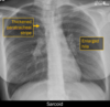
What heart abnormality might cause unilateral left hilar enlargement?

A

Pulmonary valve stenosis

3
Q

What are the chest x-ray findings of pulmonary edema?

A
  • Enlarged cardiac silhouette
  • Enlarged, ill-defined pulmonary vessels
  • Bilateral airspace opacities
  • Pleural effusions
4
Q

Where are the borders of the secondary pulmonary lobule (SPL)?

A

Interlobular septa, containing pulmonary veins and lymphatics

5
Q

What is atelectasis?

A

Collapse of airspaces.

The term encompasses mild (subsegment) to whole lung collapse

6
Q

List some common examples of lung pathologies that cause with centrilobular opacities on chest x-ray.

What structures are affected by these diseases?

A
  • Pulmonary edema
  • Bronchiolitis
  • Hypersensitivity pneumonitis

These pathologies affect the bronchioles and intralobular artery (structures in the central portion of the SPL)

7
Q

What is lobular pneumonia?

What would you see on a chest x-ray?

A

Pneumonia that encompasses an entire lobe/majority of a lobe of the lung

  • Uniform area of lung infection (no patchiness)
  • Infection is bound by a fissure
8
Q

Which structures in the hila is enlarged in sarcoidosis?

A

Lymph nodes

9
Q

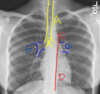
What structure is arrow #4 pointing to?

A

Pulmonary Arteries

10
Q

Describe the appearance of a pleural effusion on chest x-ray

A

Blunting of the costophrenic angles on the frontal or lateral view

11
Q

What is structure E?

A

Pulmonary Hila

12
Q

What would you see on a chest radiograph if there were a pneumonia or mass in the medial aspect of the lingula?

A

The left heart border (left ventricle border) would be obscured

13
Q

Which lobes can be thought of as the posterior lobes?

A

Lower lobes

14
Q

Which structures in the hila are enlarged in pulmonary hypertension?

A

Main and central pulmonary arteries

15
Q

What is the most likely diagnosis?

A. Pneumonia

B. Pulmonary edema

C. Lobar collapse

D. Pneumothorax

A

D. Pneumothorax

16
Q

What chest x-ray findings are you looking for when you assess the pleura?

A
  • Costophrenic angles on frontal and lateral view
    • Should be sharp; blunted in pleural effusion
    • Lateral view is more sensitive for the detection of pleural fluid
17
Q

List some common pathologies that cause with interlobular septal thickening (on CT) and Kerley B lines (on Chest x-ray).

A

Interlobular septal thickening is caused by thickening of the outer connective layer of the secondary pulmonary lobule

  • Interstitial pulmonary edema
  • Lymphatic carcinomatosis

Affect structures in the periphery of the SPL (pulmonary veins and lymphatics)

18
Q

Describe the appearance of a tension pneumothorax on a chest x-ray

A
  • Shift of the mediastinum
  • Depression of the diaphragm
  • Collapse of the lung
19
Q

In which direction is a lateral view chest x-ray taken?

A

From right to left

20
Q

How can one differentiate the interlobular septal thickening in pulmonary edema vs. lymphangitic carcinomatosis?

A

Pulmonary edema = bilateral, smooth

Lymphangitic carcinomatosis = unilateral, smooth early, nodular later on

21
Q

Which lobe(s) are collapsed?

A

Right middle lobe and right lower lobe

The right heart border and diaphragm border are obscured; caused by a lesion in the bronchus intermedius

22
Q

In a normal, healthy person, on an inspiratory frontal radiograph, the lungs extend to the ____th rib posteriorly and the ___th rib anteriorly

A

In a normal, healthy person, on an inspiratory frontal radiograph, the lungs extend to the 10 th rib posteriorly and the 6th th rib anteriorly

23
Q

List 3 key features of a normal lung x-ray

A
  • Equal density, approximately equal size
  • The bronchi and vessels are very visible centrally
  • The outer peripher of the lungs are clearer than the center
    • You should not see the peripheral bronchioles clearly
24
Q

What pathology might cause the fissures of the lungs to be seen easily?

A

Lobar pneumonia

25
Describe the appearance of a pneumothorax on a chest x-ray
Discrete visualization of the pleural line **If the patient is upright:** air collects at the apex of the hemithorax **If the patient is supine:** Air collects in the anterior, inferior hemithorax -\> **deep sulcus sign** (sharper/invaginated costophrenic angle on one side)
26
In a chest x-ray, is the PA view superior to the AP view?
The PA view more closely approximates the size of the heart AP view artificially accentuates the heart size
27
What structure is arrow #1 pointing to?
Interlobular septa contains pulmonary veins and lymphatics
28
What are the yellow arrows pointing to? What might be causing this?
Kerley B lines Caused by int**_er_**lobular septal thickening due to pulmonary edema or lymphangitic carcinomatosis
29
How many layers of pleura are in the junction lines (between lobes of the lung)?
4 One visceral and one parietal for each lobe
30
What would you see on a chest radiograph if disease were present in the lower lobes of the lung?
The diaphragm would be obscured
31
Which picture shows reticulation? Which one shows honeycombing?
* A = Reticulation * Thickened **linear** densities * Can be a sign of fibrosis * B = Honeycombing * Dilated airspaces (**rounder**), usually ~1cm * A definite sign of fibrosis
32
Which x-ray is abnormal?
A (on the left)
33
Where is the pneumonia?
Right middle lobe The right heart boarder is obscured because there is consolidation in the right middle lobe due to pneumonia
34
What pathology is shown in this chest x-ray?
Pleural effusion Blunted costophrenic angles are a classic chest x-ray finding in pleural effusion
35
What abnormalities might leftward tracheal deviation indicate?
Right aortic arch or mediastinal mass
36
What would you see on a chest radiograph if there were a pneumonia or mass in the middle lobe of the right lung?
The border between the lung and the right atrium will be obscured
37
What is the most likely diagnosis? ## Footnote A. Pulmonary edema B. Cancer C. Pneumonia D. Pneumothorax
B. Cancer Lymphangitic carcinomatosis - unilateral pulmonary edema. Fluid builds up due to blockage from tumor cells.
38
What structure is this?
Costophrenic angle
39
Which hilum is usually slightly higher?
Left hilum is usually slightly higher than the right
40
What is structure D?
Descending aorta
41
What is the CT analog for Kerley B lines on a chest radiograph? What diseases might they indicate?
CT analog = int**_er_**lobular septal thickening These findings indicate **pulmonary edema (if bilateral)** or **lymphangitic carcinomatosis (if unilateral)** Int**_er_**lobular septal thickening becomes **nodular** later on in the disease process of lymphangitic carcinomatosis (stays smooth the whole time in pulmonary edema)
42
What is an air bronchogram?
Air-filled bronchi surrounded by lung consolidation
43
What is the difference in the appearance of consolidation and ground glass opacity on chest x-ray?
Both have increased density (brighter) **Consolidation obscures** underlying structures **Ground glass preserves** underlying structures
44
List 2 diseases that could cause enlargement of the hila on chest x-ray?
* **Pulmonary hypertension:**Enlarged central pulmonary arteries * **Sarcoidosis:** enlarged hilar lymph nodes
45
What structure is this?
Left Ventricle
46
Which lobes of the lung are the anterior lobes?
Upper lobes
47
What structure is this?
Left atrium
48
What structures are at the center of the secondary pulmonary lobule?
Pulmonary veins and lymphatics
49
Which lobes of the lung touch the heart?
* Middle lobe of the right lung * Lingula of the left lung
50
What is a deep sulcus sign? What does it indicate?
Hyperluscency of the diaphragm Caused by pneumothorax in a patient who is supine; air collects in the inferior hemithorax May also see deepened, hyperluscent costophrenic angles
51
What is the smallest unit of the lung that has a connective tissue covering?
Secondary pulmonary lobule (SPL)
52
What does thickening of the interlobular septa indicate?
* Fluid in the septa (in pulmonary veins and lymphatics) * This appears as **Kerley B lines** on chest X-ray!
53
What structure is this?
Pulmonary Artery
54
When would you order a chest x-ray with an AP view?
If the patient cannot sit or stand upright Usually, you would want to ordern a PA view to minimize false magnification of the mediastinum in the chest cavity
55
What structure is arrow #2 pointing to?
Pulmonary veins + lymphatics
56
What is structure A?
Trachea
57
Which section contains "ground glass opacity?" Which section contains consolidation?
* A = Consolidation * Increased density that obscures the underlying architecture of the lung * B = Ground glass opacity * Increased density, but the underlying architecture of the lung is preserved
58
Which lung is aspirated material most likely to enter? Why?
The right lung The right mainstem bronchus is shorter and more vertically oriented than the longer, more horizontal left mainstem bronchus.
59
Where is the pneumonia?
Left lower lobe Can see the left heart border =\> not in the left upper lobe The left hemidiaphragm is obscured =\> pneumonia in the left lower lobe
60
Which fissure is the yellow arrow pointing to?
Oblique fissure, aka major fissure (this is the right lung)
61
What are paratracheal stripes?
Slightly brighter lines on either side of the trachea, where vessels and lymph nodes live They are a normal finding on a chest x-ray
62
What structure is this?
Right atrium
63
What structure is the yellow arrow pointing to?
Pulmonary vein in the interlobular septa
64
What creates a silhouette sign?
The silhouette sign is created by **differences in density**. Tehse differences allow us to see the **boarders of structures** Structures of the **same density** where **one is behind the other** will be visible Structures of the **same density** where **one is surrounding the other** will not be visible
65
What structure is arrow #3 pointing to?
Bronchioles
66
Which hilum will be enlarged in pulmonary valve stenosis?
Left hilum
67
Which 4 structures can be found in the pulmonary hila?
* Bronchi * Pulmonary arteries * Pulmonary veins * Lymph nodes
68
What causes the normal, slight-rightward deviation of the mid-thoracic trachea?
The arch of the aorta
69
Support devices like endotracheal tubes are more likely to be abnormally placed into the _[right/left]_ mainstem bronchus
Support devices like endotracheal tubes are more likely to be abnormally placed into the **_right_** mainstem bronchus
70
Where is the abnormality in this chest x-ray?
The trachea - Deviated to the left due to a right aortic arch The lungs are normal
71
What is structure B?
Carina
72
Where is the pneumonia?
Left upper lobe (lingula) Left heart boarder is obscured
73
What is structure C?
Aortic Arch
74
The segmental bronchi arise from the \_\_\_\_\_\_\_\_
The segmental bronchi arise from the **_Lobular bronchi_**