Pulm Clinical Medicine (except MDM) Flashcards

1
Q

Describe the following for Pneumothorax:

  • Percussion note:
  • Breath sounds:
  • Adventitious sounds:
  • Fremitus:
A

Pneumothorax = air in the pleural space

  • Percussion note: Hyperresonant
  • Breath sounds: Decreased or absent
  • Adventitious sounds: None
  • Fremitus: Decreased fremitus
2
Q

What conditions could cause B lines on ultrasound?

A

B lines are a marker of interstitial fluid or thickening of interstitial tissue

  • Diffuse B lines = pulmonary edema
    • Interstitial fluid
  • Focal B lines = consolidation
    • Fluid fills alveolar spaces, usually interstitial tissue is thickened
3
Q

A patient has resonant percussion, vesicular breath sounds, normal fremitus, and rhonchi.

What is most likely wrong with the patient?

A

Chronic bronchitis

Rhonchi are caused by narrowing of the large airways

4
Q

Adventitious breath sounds that are “countious, high-pitched, with a muscial quality and heard on ispiration” are most likely…

A

Stridor

5
Q

What positions of comfort might indicate that a patient is in respiratory distress?

A
  • Sniffing position
    • Indicates upper airway obstruction (this is an emergency)
  • Tripod breathing
    • Optimizes the mechanics of breathing
6
Q

Why is it important to collect “collateral information” when evaluating a patient with dyspnea?

A

Collateral information = information about changes to daily life that a patient makes to avoid dyspnea

Ex: They may resport little/no dyspnea, but what are they cutting out to avoid it?

7
Q

Adventitious breath sounds that are “continous, high-pitched, with a musical quality, and loudest on expiration with an occasional squeek” are most likely…

A

Wheezes

8
Q

What conditions would result in hyperresonance during percussion?

A
  • More air in the chest cavity (lungs or pleural space)
    • Emphysema
    • Asthma
    • Pneumothorax
9
Q

What pathological changes cause wheezes on auscultation?

A

Narrowing or partial obstruction of intrathoracic (lower) airways

May be caused by asthma, bronchitis, bronchiolitis, or airway compression

10
Q

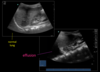
What sign is shown in this image?

What pathology does it indicate?

A

Shred sign

Indicates pneumonia

11
Q

A patient has hyperresonant percussion, decreased breath sounds, and decreased fremitus.

What is most likely wrong with the patient?

A

Pneumothorax

  • Air in the pleural space
  • Hyperresonant percussion occurs when there is increased air space in the chest
12
Q

If you hear bronchovesicular or bronchial breath sounds in abnormal places, what pathology might be present?

A

Consolidation

Due to pneumonia or pulmonary hemorrhage

In a normal lung:

  • Bronchovesicular: Large airspaces
    • 1st and 2nd interspaces anteriorly
    • Between the scapulae
  • Bronchial: Large airways
    • Over the manubrium (if at all)
13
Q

Describe the following for Pleural effusion:

  • Percussion note:
  • Breath sounds:
  • Adventitious sounds:
  • Fremitus:
A

Pleural effusion = fluid in the pleural space

  • Percussion note: Hyporesonant
  • Breath sounds: Decreased
  • Adventitious sounds: None or possible pleural rub
  • Fremitus: Decreased fremitus

Trachea may be shifted toward infolved side if a large area of the lung is affected

14
Q

In a normal lung, where would you hear bronchovesicular breath sounds? Bronchial breath sounds?

A
  • Bronchovesicular: Large airspaces
    • 1st and 2nd interspaces anteriorly
    • Between the scapulae
  • Bronchial: Large airways
    • Over the manubrium (if at all)
15
Q

A patient has resonant percussion, vesicular breath sounds, normal fremitus, and crackles.

What is most likely wrong with the patient?

A

Pulmonary edema

  • Crackles are caused by small airways popping open
  • Resonance and fremitus are normal because fluid is in the interstitium, not in the pleural space or airspaces
16
Q

What will you see on a normal ultrasound of the lung?

A
  • Bright white, horizontally-sliding pleura
  • A-lines (green)
  • Comet tails (a few)
17
Q

What will you see on ultrasound if the patient has a pneumothorax?

A
  • Comet tails are abent
  • No pleural sliding where the pneumothorax is
    • The rest of the pleura will slide
  • Lung point may be visible
    • Boundary between normal lung and pneumothorax
18
Q

What conditions would result in increased tactile fremitus?

A
  • Things that increase the amount of fluid in the lungs
    • Pneumonia
    • Pulmonary hemorrhage
19
Q

If a patient is in respiratory distress, what signs can be seen in the neck?

A

Trapezius and sternocleidomastoid contraction

Tracheal tugging

20
Q

What special tests can be performed to assess for consolidation of the lungs?

A
  • Bronchophony
    • Words will be louder than normal - “99”
  • Whispered Pectoriloquy
    • Intesnsification of whispered words - “1, 2, 3”
  • Egophony
    • Normal “ee” sounds will sound like a long “a”
21
Q

If you hear stridor, what pathology is most likely present in the patient?

A

Upper airway obstruction

  • Croup
  • Laryngeal edema
  • Airway compression
  • Airway stenosis
22
Q

What are the 4 key inputs that can drive dyspnea?

A
  • Mechanoreceptors
  • Chemoreceptors
  • Neurohormonal uncoupling
  • Psychosocial factors

Usually dyspnea is a combination of several of these factors

23
Q

A patient presents with a cough. List the alarm sysmptoms that would warrant expedited or urgent evaluation

A

CHeWW-D CHESS

  • Current/Former smoker with a New Cough
  • Hemoptysis
  • Wheezing and shortness of breath
  • Weight gain
  • Nocturnal Dyspnea
  • Chest pain
  • Hoarseness
  • Peripheral Edema
  • Trouble Swallowing
  • Systemic (fever, weight loss)
24
Q

Describe vesicular breath sounds

A

Normal breath sounds

  • Heard over most of the lung field (except near large airways)
  • Louder in inspiration than expiration
25
If a patient is working harder than usual to breathe, what facial signs might be present?
* Nasal flaring * Head bobbing * Retractions * Grunting/breath holding * Neck muscle use
26
What pathological changes cause stridor on auscultation?
Narrowing of the **extrathoracic** (upper) airway May be due to croup, laryngeal edema, mass
27
On which side of the chest is it important to listen in the mid-axillary line?
Right You are listening for the right middle lobe (the left lung does not have a middle lobe)
28
Describe the following for **Consolidation**: * Percussion note: * Breath sounds: * Adventitious sounds: * Fremitus:
Consolidation = fluid in the alveolar spaces * Percussion note: **Hyporesonant** * Breath sounds: **Decreased** * Adventitious sounds: **Crackles (late inspiratory)** * Fremitus: **Increased fremitus**
29
A 68 year-old man has severe smoking-related chronic obstructive pulmonary disease (COPD) causing lung hyperinflation. He takes an inhaled corticosteroid, an inhaled long-acting beta agonist, and an inhaled long-acting muscarinic antagonist daily. He uses 2 liters per minute supplemental oxygen to maintain adequate oxygen saturations at rest and with activity. The patient reports significant dyspnea with activity. The _least_ _likely_ cause of this patient’s dyspnea is: 1. Hypoxemia 2. Lung hyperinflation 3. Deconditioning 4. Severe airflow obstruction
a. Hypoxemia Hypoxemia does not have a strong association with dyspnea. Additionally, lung hyperinflaiton, deconditioning, and sever airflow obstruction are all commonly seen in patients with COPD
30
Adventitious breath sounds that are "brief, intermittent, and discontinuous" are most likely...
Crackles
31
What pathological changes cause rhonchi on auscultation?
Narrowing of **larger** airways from secretions May be present in bronchitis, bronchiolitis, COPD, and asthma
32
What are the 4 most important questions that must be answered when a patient presents to your clinic with a cough? (You don't need to ask the patients these exact questions, but you need them answered in order to make a diagnosis)
1. Is the cough acute or chronic? 2. Are any alarm symptoms present? 3. Does the patient have comorbid conditions (COPD, asthma, bronchiectasis, rhinosinusitis, GERD) 4. Is environment or medication playing a role?
33
Will a patient wtih asthma have increased fremitus or decreased fremitus? Why?
**Decresed** Asthma is an obstructive lung disease, resulting in hyperinflation of the alveoli. Fremitus is decreased when there is increased air in the alveolar spaces
34
If your patient has pneumonia, what will you see on ultrasound?
* Focal B lines * Chains of white dots, indicate lung consolidatiosn * Lung hepatization * Dynamic air bronchograms * Shred sign * Disruption of the pleural line, like a bite is taken out
35
What is the differential diagnoses for wheezes?
Asthma Bronchitis Bronchiolitis Airway compression (foreign body or mass)
36
What conditions would result in decreased tactile fremitus?
* Things that increase **air or fluid** in the **pleural space** * Pleural effusion * Pneumothorax * Things that increase **air** in the **lung** * Asthma * Emphysema
37
List the 2 primary causes of neurohormonal uncoupling that contribute to dyspnea
1. Increased load on the respiratory system - Asthma, COPD, pneumonia, pulm. edema, ILD 2. Respiratory muscle weakness - Muscle fatigue, neuromuscular weakness/disease, lung hyperinflation (associated with COPD)
38
What is the differential diagnosis for rhonchi?
Bronchitis Bronchiolitis Asthma
39
Which ultrasound probe has the highest resolution? Which one penetrates the deepest?
Highest resolution = high frequency probe Deepest penetration = low frequency probe
40
What are the 5 major goals in treating patients with dyspnea?
1. Treat the underlying cause - Bronchodilators, drain pleural effusion, treat anemia 2. Increase respiratory muscle function - Pulmonary rehabilitation, nutrition 3. Decrease chemoreceptor input - Give oxygen, noninvasive ventilation 4. Decrease central respiratory drive - Opioids in palliative care 5. Education and coping - Pacing, cool air, pursed-lip breathing
41
What will you see on ultrasound if the paient has a pleural effusion?
* Spine sign * Fluid surrounding the lung allows ultrasound waves to penetrate, we can then visualize the vertebral bodies * Dark triangle of fluid
42
Describe the following for **Upper Airway Obstruction**: * Percussion note: * Breath sounds: * Adventitious sounds: * Fremitus:
Upper Airway Obstruction = extrathoracic; croup, laryngeal edema, laryngomalacia * Percussion note: **Resonant** * Breath sounds: **Vesicular, decreased if obstruction is severe** * Adventitious sounds: **Stridor** * Fremitus: **Normal fremitus**
43
Describe the following for **Bronchiolitis**: * Percussion note: * Breath sounds: * Adventitious sounds: * Fremitus:
Bronchiolitis = narrowing of the lower (intrathoracic) airway * Percussion note: **Resonant** * Breath sounds: **Obscured by adventitious sounds** * Adventitious sounds: **Wheezing, rhonchi, crackles** * Fremitus: **Normal fremitus**
44
Describe the following for **Asthma**: * Percussion note: * Breath sounds: * Adventitious sounds: * Fremitus:
Asthma = Hyperinlated alveoli * Percussion note: **Resonant to hyperresonant** * Breath sounds: **Often obscured by adventitious sounds** * Adventitious sounds: **Wheeze,** possible crackles & rhonchi * Fremitus: **Decreased fremitus**
45
Which ultrasound is abnormal? What is the pathology?
There is a pneumothorax in the image on the left
46
What will you see on ultrasound if the patient has edema?
* Many B lines (diffuse) * Bright white lines that shoot all the way down to the bottom of the screen from the pleura * More than a few = abnormal * They move back and forth with respiration as the pleural line moves * **More B lines =\> more fluid in the pleura** * If severe, the B lines may coalesce to obscure A lines
47
What is the differential diagnosis for crackles?
Fibrosis Pneumonia Pulmonary edema
48
List 4 locations for mechanoreceptors that are important in sensing dyspnea
1. Face/upper airways 2. Pulmonary stretch receptors 3. Irritant receptors 4. Chest wall
49
A patient has hyporesonant percussion, decreased breath sounds, and decreased fremitus. What is most likely wrong with the patient?
Pleural effusion Decreased fremitus when there is fluid **outside** of the alveolar spaces
50
What comorbidities are important to look out for when you are evaluating a patient for cough?
COPD Asthma Brochiectasis Rhinosinusitis GERD
51
What pathology does this lung show?
Consolidation May be caused by pneumonia, alveolar hemorrhage, or anything that causes fluid to replace air in the alveolar spaces
52
What is the differential diagnosis for stridor?
Stenosis of the large airway Laryngeal Edema Compression Croup
53
Which image is abnormal? How do you know?
The image on the bottom/right is abnormal. The black triangle and spine sign indicate a **pleural effusion** Spine sign = you can see the spine clearly Black triangle = fluid in the pleural space
54
What pathological changes cause crackles on ascultation?
Small airways popping open during inspiration (May be related to secretions in closed, small airways) Usually due to fibrosis, pneumonia, or pulmonary edema
55
Any cough lasting **more than ____ weeks** is considered chronic
Any cough lasting **more than _8_** **weeks** is considered chronic
56
Any cough lasting **less than ____ weeks** is considered acute
Any cough lasting **less than _3_ weeks** is considered acute
57
Describe bronchovesicular breath sounds
Heard over large airspaces * Inspiratory and expiratory components are equal in volume * In a normal lung: * 1st and 2nd interspaces anteriorly * Between the scapulae
58
A normal adult respiratory rate is ______ breaths/minute
A normal adult respiratory rate is **_10-14_** breaths/minute
59
Describe the following for **Emphysema**: * Percussion note: * Breath sounds: * Adventitious sounds: * Fremitus:
Emphysema is a pathology of the alveoli * Percussion note: **Hyperresonant** * Breath sounds: **Decreased** * Adventitious sounds: **None** * Fremitus: **Decreased fremitus**
60
A 35-year-old male presents to the emergency room for 2 weeks of coughing. He is a non-smoker, has no known medical problems, and denies any alarm symptoms. His vital signs and physical examination are unrevealing. A chest radiograph is normal. Which is of the following is the appropriate next step? 1. Chest CT 2. Cough-suppressant therapy with outpatient follow-up in 4 weeks 3. Bronchoscopy 4. 5-day course of antibiotic therapy for community acquired pneumonia
B. Cough-suppressant therapy with outpatient follow-up in 4 weeks Acute cough + reassuring physical examination + normal x-ray = probably a viral respiratory tract infection that can be managed conservatively with close follow-up
61
Adventitious breath sounds that are "continous, with a low-pitched, sonorous, musical quality" are most likely...
Rhonchi
62
What conditions would result in hyporesonance during percussion?
* Increased fluid in the chest cavity * Consolidation (Fluid in alveolar spaces) * Atelectasis * Pleural effusion
63
A 34 year-old woman presents with worsening breathlessness over the past 3 days. She describes a sensation of “chest tightness.” The most likely diagnosis is: 1. Pneumonia 2. Pneumothorax 3. Asthma exacerbation 4. Pulmonary embolism
c. Asthma exacerbation "Chest tighness" to describe dyspnea is commonly associated with acute bronchospasm, seen in asthma or COPD exacerbation
64
If a patient is in respiratory distress, what signs might be present in the chest/abdomen?
Intercostal or subcostal retractions Accentuated abdominal breathing
65
When imaging the chest, when would you use a high-frequency ultrasound probe?
When you are evaluting the pleural line or looking for a pneumothorax High frequency probes penetrate more shallowly but have higher resolution
66
List the order of step-wise testing and treatment of a chronic cough
One at a time! 1. **Asthma** - Try corticosteroid therapy 2. **GERD** - Try acid-suppression therapy 3. **Rhinosinusits** - Try nasal corticosteroids + antihistamine therapy
67
Describe the following for **Pulmonary Edema**: * Percussion note: * Breath sounds: * Adventitious sounds: * Fremitus:
Pulmonary edema = fluid in the interstitium * Percussion note: **Resonant** * Breath sounds: **Vesicular** * Adventitious sounds: **Late inspiratory crackles,** possible wheezing * Fremitus: **Normal fremitus**
68
Describe the following for **Atelectasis**: * Percussion note: * Breath sounds: * Adventitious sounds: * Fremitus:
Atelectasis = lobular obstruction from mass, mucus, or foreign object * Percussion note: **Hyporesonant** * Breath sounds: **Decreased or absent** * Adventitious sounds: **None** * Fremitus: **Normal fremitus**
69
What are the three most common causes of a chronic cough?
Asthma GERD Rhinosinusitis
70
Describe the following for **chronic bronchitis**: * Percussion note: * Breath sounds: * Adventitious sounds: * Fremitus:
Chronic bronchitis is a pathology of the upper airway * Percussion note: **Resonant** * Breath sounds: **Vesicular** * Adventitious sounds: **Rhonchi,** crackles, wheezes * Fremitus: **Normal fremitus**
71
Which measurement contributes most significantly to a sensation of dyspnea? 1. Low PaO2 2. High PaCO2 3. Low pH 4. High pH
B. High PaCO2 Increased PaCO2 = Hypercapnia Induces dyspnea in the absence of respiratory muscle activity
72
Describe bronchial breath sounds
Heard over large airways * Breath sounds are louder and more high-pitched * Expiratory component is louder than inspiratory * Heard over the manubrium (if at all) in a normal lung
73
On ultrasound, fluid appears ______ and air appears \_\_\_\_\_\_\_.
On ultrasound, fluid appears **_black**_ and air appears _**white._**
74
A 65-year-old male presents to urgent care for evaluation of a cough which has been present for 10 weeks. He has smoked 1 pack of cigarettes per day for 40 years. He notes his sputum is frequently streaked with blood and that he has lost 15 pounds in the last 2 months. His vital signs are unremarkable and he has no focal findings on physical exam. The appropriate next step in management is... 1. Arrange for a non-urgent chest radiograph and prescribe a cough suppressant 2. Trial of an inhaled corticosteroid for possible cough-variant asthma 3. Urgent chest radiograph, complete blood count, and chemistry panel 4. Prescribe a 5-day course of azithromycin for possible bacterial pneumonia
C. Urgent chest radiograph, complete blood count, and chemistry panel Alarm symptoms are present (new cough in an active smoker, hemoptysis, weight loss). Evaluate urgently for malignancy
75
When imaging the chest, when would you use a low-frequency ultrasound probe?
* When you are looking for pleural effusion or hemothorax * When the patient is larger The low-frequency probe penetrates deeper but has lower resolution
76
77
What is the difference between wheezes and stridor? In which disease processes would you hear each?
* Wheezes * Ususally expiratory * Caused by narrowing of the **int****rathoracic (lower) airway** * Seen in asthma, bronchiolitis, bronchitis, and airway compression * Stridor * Usually inspiratory * May contain squeeks * Caused by narrowing of the **extrathoracic (upper) airway** * Seen in airway stenosis, laryngeal edema, compression, croup
78
What is the difference between crackles and rhonchi? In which disease processes would you hear each?
* Crackles * Discontinuous * Can be fine and dry or coarse and wet * Heard in fibrosis, pneumonia, and pulmonary edema * Rhonchi * Continuous - Usually inspiratory and expiratory * Low-pitched snoring * Heard in bronchitis, asthma, and bronchiolitis
79
A patient has hyporesonant percussion, decreased breath sounds, increased fremitus, and crackles. What is most likely wrong with the patient?
**Consolidation** * Fluid in the alveolar spaces, likely caused by pneumonia or hemorrhage * **Increased fremitus** occurs when there is fluid inside​ of the alveolar spaces * **Hyporesonant** precussion occurs when there is fluid, consolidation, or mass in the chest cavity
80
A 34 year-old man with hereditary hemorrhagic telangiectasia presents with worsening dyspnea on exertion of 3 month’s duration. On exam, he appears pale but in no acute distress. Vital signs are normal except for a resting heart rate of 105 beats per minute. Examination demonstrates lip telangiectasias, normal heart tones, and normal breath sounds. His oxygen saturation on room air at rest is normal. The most likely cause of this patient’s dyspnea is: 1. Pulmonary hemorrhage 2. Anemia 3. Myocardial ischemia 4. Pulmonary embolism
b. Anemia Anemia is linked to chronic dyspnea; the other options all cause acute dyspnea
81
Dyspnea that is described as "chest tightness" has a high association with \_\_\_\_\_\_\_\_\_\_\_\_\_\_\_\_
Dyspnea that is described as "chest tightness" has a high association with **_bonchospasm caused by Asthma or COPD_**
82
What are the advantages of chest CT over chest x-ray?
In a chest CT... * No blind spots * Can reconstruct in multiple dimensions
83
What are the disadvantages of CT compared to chest x-ray?
Hight cost Higher radiation dose
84
What are the indications for IV contrast in a CT?
* Evaluation of vasculature * Dissection (CT angiogram) * Pulmonary embolism (CTPA) * Evaluation of solid organs * Evaluation of mediastinum * Lung cancer **staging** (Not screening) Contrast not needed to look for lung nodules in screening!
85
What kind of imaging would you use to evaluate a pulmonary embolism?
CT with IV contrast
86
What kind of imaging is used for lung cancer screening?
Low dose CT | (No contrast)
87
Low-dose chest CT is about the same radiation dose as ______ chest x-rays Routine chest CT is about the same radiation dose as \_\_\_\_\_\_ chest x-rays
Low-dose chest CT is about the same radiation dose as **_10-15_** chest x-rays Routine chest CT is about the same radiation dose as ​ **_50_** chest x-rays
88
NLST showed a \_\_\_\_\_% mortality benefit in chest CT screening group compared wtih the chest x-ray screening group
NLST showed a **_20%_** mortality benefit in chest CT screening group compared wtih the chest x-ray screening group
89
What are the indications for lung cancer screening? Age: Smoking history: Symptoms:
Age: **55-77 years old** Smoking history: **\>30 pack years, quit \<15 years ago** Symptoms: **Asymptomatic**
90
What is the major contraindication to IV iodinated contrast?
Renal impairment
91
Where are the "blind spots" on a chest x-ray? How can we visualise them?
Behind the diaphragm Behind the mediastinum In the lung apices Visualize with chest CT
92
What kind of imaging is used in lung cancer staging?
Routine chest CT with contrast
93
What kind of imaging is used to evaluate interstitial lung disease?
HRCT
94
Radiation doses in various imaging modalities A chest radiograph is about ____ mSv A low-dose chest CT is about _____ mSv A regular chest CT is about _____ mSv
A chest radiograph is about **_0.1_** mSv A low-dose chest CT is about **_1-2_** mSv A regular chest CT is about **_7_** mSv ​Being alive for 1 year ~2 mSv
95
What is the most likely diagnosis of a 4 mm lung nodule in a smoker with \>30 pack year history?
Granuloma Very common! If nodule size has not changed in 1 year =\> granuloma and not cancer (biopsy not necessary)
96
What are the most common harms of lung cancer screening?
* Additional testing due to incidental findings * Ex: Biopsy of a granuloma * Overdiagnosis * Ex: finding a carcinoid tumor that would not have impacted the patient's life
97
If disease prevalence is **low**... Positive predictive value will be [low/high] and Negative predictive value will be [low/high]
If disease prevalence is **low**... Positive predictive value will be **_low_** and Negative predictive value will be **_high_**
98
What is is the first line imaging test for the diagnosis of pneumonia or other common disease processes?
Chest x-ray Chest CT can be performed if cxr is negative but clinical suspicion is high