SI Flashcards

1
Q

When does chyme move into the stomach

A

0-3 hrs after entering the stomach

2
Q

How long does it take for chyme to move through the SI (6m)

A

1-6 hours

3
Q

Role of

  1. Duodenum
  2. Jejunum
  3. Ileum

in absorption

A
  1. Digestion and a small amount of absorption
  2. Digestion and absorption
  3. Absorption
4
Q

Conduction rate of the SI

A

1 cm/min

5
Q

What is the conduction rate of the SI influenced by

A
  • Enteric nervous system
  • Hormones
  • Gastrin (increase)
  • Cholecystokinin (increase)
  • Secretin (increase)
6
Q

Relationship between structure and SA of the SI

A
7
Q

Actual SA of the SI

A

120-140 m2

> 95% of nutrients are absorbed

Increasing food intake, limited capacity - reserve capacity (when we over-eat)

8
Q

Structure of SI

A

ENS regulates digestion, motility and absorption

9
Q

Gut layer organisation

A

Gut wall has a layered organisation, with the absorptive cells lining the lumen and neural muscular components below

Blood and lymph vasculature is abundant to transport absorbed nutrients

10
Q

Function of villi

A

By projecting into the lumen, the villi increase the SA for absorption of nutrients

Microvilli (brush border) fringe the villi to further increase SA

11
Q

What does lymph carry

A

Fat and fat-soluble vitamins

12
Q

Structure of SI

A
13
Q

What is the epithelial mucosa composed of

A

Absorptive cells

Secretory cells - enzymes, hormones, fluid (follows secretion of solutes), mucus (for protection)

14
Q

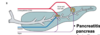
What do the exocrine cells in the pancreas do

A

Play a central role in the production of digestive enzymes

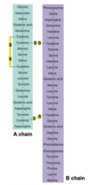
(beta cells - islets of langerhans - secrete insulin into circulation)

15
Q

Where are digestive secretions from the liver and pancreas delivered into

A

Into the duodenum through the SPHINCTER OF ODDI

16
Q
  1. Acini secrete…
  2. What stimulates acini
A
  1. Enzymes
  2. CCK and ACh
17
Q
  1. Ducts secrete…
  2. What are the ducts stimulated by
A
  1. Bicarbonate
  2. Secretin
18
Q

What does CCK do

A

Makes gallbladder contract

CCK mediates gastric emptying rate in response to fat

Reduces appetite and induces nausea

19
Q

What protects the pancreas from autodigestion

A

Kazal inhibitor secreted from pancreas to protect it

20
Q

Bicarbonate secretion in pancreas or intestine

A

(Position of transporters is reversed in the stomach)

  • BASOLATERAL - Na+ will be used to export H+ out of the cell into circulation
  • H+ came from carbonic anhydrase that broke down from CO2 and H2O
  • Bicarbonate is exchanged for chloride on the luminal side of pancreas - Cl- can flow back in
  • Chloride is circulating across
  • NET EFFECT: Bicarbonate into lumen - sodium goes between cells and leaks into lumen, dragging water by osmotic load
21
Q

What does the stomach have (in relation to HCO3- secretion)

A

Stomach has defensive mucus layer filled with continuous secretion of bicarbonate, so the mucus layer in the stomach protects the gastric wall from being attacked by acid

We must neutralise acidic chyme to prevent intestinal ulcers

Also neutral pH for enzymes to act

22
Q

What is secreted from the pancreas

How is it secreted

A
  • NaHCO3- solution
  • Cl-/HCO3- antiport co-transporter on apical membrane
  • HCO3- enters pancreatic duct
  • Na+ secreted through cell junctions
23
Q

How else is NaHCO3 secreted, apart from the pancreas

A

By liver (bile)

24
Q

Response of an increase in acid from the stomach

A
  • Sensors in duodenum
  • Secretin conc will rise
  • Stimulate pancreas to release bicarbonate, bicarbonate will flow into SI, neutralise acid and reduce the stimulus on secreting cells
  • -ve feedback loop controls HCO3-
  • Sensor picks up pH of SI
  • Acid is neutralised, SI protected
25
Where are secretin's receptors
Found in the pancreas, which responds with additional bicarbonate delivery Gastric motility and secretion are inhibited
26
TRYPSIN, CHYMOTRYPSIN, ELASTASE 1. Substrate 2. Action
1. Protein 2. Breaks peptide bonds in proteins to form peptide fragments 5-10 AAs long
27
CARBOXYPEPTIDASE 1. Substrate 2. Action
1. Proteins 2. Splits off terminal AA from carboxyl end of protein
28
LIPASE 1. Substrate 2. Action
1. Fats 2. Splits off 2 FAs from TAGs, forming free FAs and monoglycerides Secretion of lingual lipase in saliva is MINOR - mostly lipases from pancreas split FAs from TAGs
29
AMYLASE 1. Substrate 2. Action
1. Polysaccharides 2. Splits polysaccharides into glucose and maltose
30
RIBONUCLEASE, DEOXYRIBONUCLEASE 1. Substrate 2. Action
1. Nucleic acids 2. Splits nucleic acids into free mononucleotides Digestive enzymes isn't just about breakdown of building blocks into smaller ones - causing amnesia in food - loses its original aim/design and become completely inert building blocks Coronavirus will have RNA in it - injected into cells and our mechanisms will express the proteins of the coronavirus rather than our own RNA, which is important because it will break down external RNA Ribonucleases are important, not just to bring energy into the cell or generate BBs, but protect us too
31
Examples of carbohydrates in foods
32
What is the importance of glucose homeostasis i.e. [Glu]plasma
Major fuel for brain function
33
Food energy sources
* Fat * Protein * CHOs * Alcohol * Water * Vitamins * Minerals
34
Rule of 2
* Survive 2 mins without O2 * Survive 2 days without water * 2 months without food * 2 years without vitamins and minerals
35
How much CHO do we use a day How much of this does our brain use
250g 125g CHO intake does not change much over the years
36
Glucose usage in muscle and adipose tissue
* Major end target for glucose is muscle - largest mass of tissue in the body * Adipose tissue can take some glucose in * Adipose tissue and muscle are INSULIN DEPENDENT (some glucose uptake in muscle is INdependent)
37
Glucose uptake in brain
Insulin INDEPENDENT
38
When might we lose glucose from plasma into urine
If plasma conc is high as glucose will not be absorbed quickly enough in the PCT
39
What is normal plasma glucose conc
Plasma glucose conc is 5mM (which in 3L of plasma is 6g of glucose and yet we'll push 250g across a small compartment - turnover rate will change dramatically) fasting and doesn't rise above 10 mM in healthy
40
Schematic representing glucose absorption
41
CHO digestion
Oligosaccharides (4-10 monosaccharides)
42
Examples of CHOs
43
What is starch
The way plants store CHOs
44
What is glycogen
The way animals store CHOs
45
Digestion of CHOs
Digestion in lumen of starches into shorter and shorter bonds until we have disaccharides floating the lumen Finally digested on brush border membrane by disaccharidases (e.g. sucrose -\> glucose and fructose in the presence of sucrase)
46
Lactase deficiency
Inability to digest lactose, ends up in LI, bacteria in LI feast and produce gas and toxins
47
CHO absorption
* Na+ gradient couples sugar absorption into cell * 2 sodium ions in, 1 glucose in also * Chloride is meant to be transported both through the cell and between cells * Net effect - sodium glucose absorption, ends up in blood stream, dragging water by osmotic load
48
Challenges in CHO absorption
Glucose, galactose and fructose are all polar molecules SGLT1 imports sodium and glucose Glucose will go through the GLUT 2 transporter which facilitates diffusion on the basolateral side of the membrane, into the bloodstream
49
What was SGLT1 predicted to be
A transmembrane protein
50
What does this graph show
SGLT1 does not account for all the absorption of glucose At 50 mM it peaks and there's no further increase in its rate of glucose absorption GLUT2 into apical membrane that is responsible for the high concentrations
51
Conc of glucose in experiments
* 1 M conc of glucose has 180g of glucose in 1 L of water * Over the years, experiments have used 100 - 800 mM of glucose * Conc have gone down to 50-300 mM * No more than 100 mM in SI * Proximal SI - 100-150 mM is reasonable * Distal SI (jejunum and ileum) - conc drop - 1-2 mM * LI - 0.5 mM or less * Conc in plasma - 7-10 mM (in rats) * Secrete more into stomach to dilute content * Keep experimental data relevant to humans - testing at 800 mM is not relevant
52
How are most CHOs in the diet consumed
As disaccharides or polysaccharides
53
Name the major disaccharides and polysaccharides
DISACCHARIDES: * Sucrose * Lactose * Maltose POLYSACCHARIDES: * Starch * Glycogen * Cellulose (only in LI as we do not have the enzymes to break the glucose glucose bonds in cellulose, so bacteria in LI is used to digest some of these fibres)
54
CHO absorption
* Brush border enzymes are attached to the luminal surface of enterocytes * Disaccharides -\> monosaccharides - Fructose - Glucose - Galactose * SGLT1 will transport either glucose or galactose against a conc gradient * Na+ gradient generated * Glucose is being absorbed - for glycogen, make proteins out of it, oxidise and get ATP
55
Another function of CHO absorption
Any bacteria still in chyme are exposed to a low glucose conc - glucose is produced directly next to glucose transporters - brush border membrane area that generates glucose and quickly absorbs it, keeping conc of glucose in the lumen low - also a feature of protecting the body from bacterial infection as bacteria also like glucose - enters glycolysis, generates energy for any cell very quickly so we keep conc of glucose in lumen low by having disaccharides on the brush border enzyme and not floating around inside lumen
56
What is different about the digestion of protein
Site of the end of digestion is NOT in the lumen
57
What is this structure What do the sulfur bonds provide
Insulin S bonds provide stability
58
What would be the effect if I expressed insulin in an artificial solution and drank it - would plasma glucose change
* Plasma glucose would not change as insulin is a protein - it will be digested in the stomach by pepsin and then trypsin in the SI - AAs will then be absorbed * In order to act as insulin it must retain its structure - injected into venous system
59
What would happen if digestive enzymes were synthesised in their active form
They would digest the very cells that make them HENCE inactive precursors (e.g. trypsinogen) become activated (trypsin)
60
How does trypsinogen become trypsin
* Enterokinase is stuck onto the brush border - luminal side * Cleave some Aas off trypsinogen and turn it into trypsin * \*\* in the stomach pepsin was activated by pepsinogen and acid
61
Where are CCK's receptors located
* In the pancreas, which responds with additional enzyme delivery (activates through 2nd messenger) * In the gallbladder, which contracts to deliver more bile * In the sphincter of Oddi, which relaxes to facilitate delivery of the enzymes and bile salts
62
What is the major stimulus for the release of CCK
FAs and AAs are sensed in the duodenum
63
-ve feedback loop of CCK secretion
Digestion and absorption of FAs reduces the signal and at the end of the meal we will end this digestion phase and SI will be empty
64
Pancreatic enzymes (PROTEASES) involved in digestion of proteins
Trypsin, chymotrypsin * Digest proteins to peptides * Trypsin activates other proteases, peptidases * Trypsin inhibitor secreted by pancreas to block action - KAZAL FACTOR adheres to trypsin and prevents it from being active
65
Pancreatic enzymes (CARBOXYPOLYPEPTIDASES) involved in digestion of proteins
* Digest polypeptides to small peptides and AAs * Carboxy-terminal AA
66
Brush border proteases
ENTEROKINASE Activates trypsin
67
Brush border peptidases
AMINOPOLYPEPTIDASES * Digest polypeptides to small peptides and AAs * Amino-terminal FUNCTION 1. Digestion for purpose of absorption 2. Helps to keep bacteria under control
68
How are free AAs, mono, di and tri-peptides absorbed \*UNUSUAL\*
* Absorbed into epithelium by active transport/facilitated diffusion, mainly in crypts (Na+ co-transport mechanism similar to glucose - SGLT1) * Digested inside the enterocytes - site where digestion ends is inside the enterocyte (epithelial cell)
69
What do cytoplasmic peptidases do
Digest peptides to AAs
70
How are some small peptides carried across the epithelium
By transcytosis
71
Antigens involved in protein absorption
* Development of food allergies and intolerances (e.g. gluten, coeliac disease) * Autism, schizophrenia, Parkinson's disease * Prion disease * Potential for development of indigestible peptide drugs (e.g. peptide hormone replacement therapy)
72
Protein digestion overview
* Proteins are broken down to peptide fragments in the stomach by pepsin, and in the SI by trypsin and chymotrypsin, the major proteases secreted by the pancreas * These fragments are further digested to free AAs by carboxypeptidase from the pancreas and aminopeptidase, located on the luminal membranes of the SI epithelial cells * The free AAs then enter the epithelial cells by secondary active transport coupled to Na+ * Short chains of 2 or 3 AAs are also absorbed by a secondary active transport coupled to the H+ gradient
73
Movement of peptides across lumen, intestinal epithelial cell and IS fluid
AAs leave by faciliated diffusion and get carried away through the portal vein into the liver
74
Why does lipid digestion take longer than other nutrients
* Takes longer than all the other nutrients * Lipids are not soluble in water * Most of our proteins are water soluble material * Lipid environment requires emulsification * Full of fat, but lots of it not available for digestion or absorption * Must access fat before we can digest it - long delay
75
What is essential for emulsification
Bile salts
76
CCK and lipid digestion
* Bile salts are secreted by the liver, stored in gallbladder * in response to CCK gallbladder will contract and release bile salts into duodenum because CCK also relaxes the sphincter of Oddi
77
What is bile secreted by
Hepatocytes
78
Contents of bile
* Cholesterol * Bile salts - cholesterol and AAs * Lecithin (phospholipid) * Bile pigments - bilirubin, biliverdin * Water * NaHCO3
79
Max capacity of the gallbladder
60 ml
80
How can bile be concentrated
By absorption of fluid and electrolytes by epithelium of gallbladder
81
Trigger of the release of bile
CCK ACh Sphincter of Oddi relaxes
82
What are gall stones
Crystals of cholesterol High fat diet and inflammation of mucosa
83
Function of bile - DIGESTION
EMULSIFICATION * Bile salts and lecithin * Mechanical processes break up fat droplets * Lipases digests fats in the presence of colipase - a cofactor needed for lipase to penetrate bile salts
84
Function of bile - ABSORPTION
MICELLE FORMATION * Complexes with a sterol nucleus that ferrys fats through the brush border to allow absorption into epithelial cells * Bile salts reabsorbed in ileum - 94% * Re-secreted
85
Structure of bile salt
Non-polar surface helps to emulsify fats Polar surface promotes water solubility
86
MOA of bile salts
* Biphasic molecule - bile salts can attach to fat molecules with its fat soluble side and attach to water with its polar side * =\> EMULSIFY FAT * Emulsify is the bringing together of fat and water in very close approximation * Making a very well mixed solution of fat * We're really increasing the surface area * Imagine a football full of marbles - Skin is suitable to aqueous environment, but when marbles are allowed out SA increases massively (emulsification) * SA:vol ratio is INCREASED - making it accessible to our water soluble enzymes * Phospholipids can help - every cell membrane is composed of phospholipids - they too have a polar end and a fat soluble end * Importance of CHEWING - breaking up cell membranes - maybe the function is to facilitate emulsification of fat - so fat can be sensed in the duodenum * +VE FEEDBACK LOOP - release of phospholipids will facilitate emulsification - facilitate digestion in SI
87
What happens once the fat globule has been broken down into smaller pieces
* Lipases from pancreatic origin will digest the emulsified fat * TAGs = 95% * LCFAs - 16 or 18 Cs * Lipases cleave off/hydrolyse the FAs off the TAG and take position 1 and 3 off * Molecules must migrate across the mucus layer * Cannot readily diffuse across the mucus layer - form micelles, spheres with water soluble component of the molecule facing out and fat soluble component inside * These micelles will migrate across the brush border membrane and end up adjacent to the epithelial cell * Diffusion does NOT happen - fat is an energy dense nutrient * Diffusion suggests that when conc of FAs is low in the lumen, FAs can just diffuse back out - extremely unlikely that we'd just lose this nutrient after we used so much energy to acquire and digest food
88
Overview of fat digestion
* NOT DIFFUSION but at least 3 classes of specified transporters that transport Fas into the cell * End of digestion by pancreatic lipases and absorption of Fs is TRANSPORTER mediated * Inside the cell we re-synthesise TAG and re-package them * Enzymes inside enterocytes that will take Fas and monoacylglycerols and form a TAG again * ER will package those cells - CHYLOMICRON, which has an apolipoprotein (specialised protein in enterocytes that allows binding of lipids, together with some cholesterol) * Chylomicron has a water-soluble outside, lipid soluble core - way to transport fats in the bloodstream - we can't have free FAs in the bloodstream as they'll just float to the top and damage the brain * We must have similar density molecules and chylomicrons serve that function * In systemic circulation chylomicrons will be re-circulated so they can feed tissues or else stored in adipocytes
89
Structures in this image
* Microvilli at the top * Middle cell - nucleus - chromatin (DNA) around the rim * Secreted on basolateral side - rate-limiting step for absorption of lipids is the ability of cell to export chylomicrons into the left
90
How are the fat soluble vitamins absorbed
* Like other lipids * Move freely with lipid phase * Taken up by diffusion
91
How are water-soluble vitamins absorbed
By diffusion or mediated transport, except for VITAMIN B12, which must first bind to a transport protein known as intrinsic factor (secreted by the stomach)
92
Summary of liver functions
93
Water homeostasis
* Secrete into lumen and then reabsorb in the lumen * Water secretion make up 6L a day * We reabsorb about 7 L daily * Much greater absorptive capacity than we'd imagine * 1L drinking * 0.5L from food * Reabsorbed in SI by osmosis * Osmotic drag - across epithelium into IS fluid and then plasma * What moves through? 1.5L of water, some fibre that wasn't digested or absorbed, some cells shed from villi * Low energy density moves to LI