SCRUBS work Flashcards
ligaments of wrist
radial and ulnar collateral ligaments -> limit ulnar and radial deviation
palmar radiocarpal -> limit extension
dorsal radiocarpal -> limit flexion
Intercarpal and carpometacarpal -> intrinsic
palmaris brevis
ulnar nerve
protects ulnar artery and nerve
deepens cup of palm

wrist frxs
Distal radius/ulna:
- Colles’ frx: hyperexternsion, fall on oustretched hand, dinner fork deformity, distal fragment displaced posteriorly w/ posterior tilt
- Smith’s/Reverse Colles’ frx: hyperflexion, garden spade deformity, distal fragment displaced anteriorly w/ anterior tilt
- scaphoid: fall on outstretched hand, frx across waist of bone, pain in anatomical snuff box, difficult to diagnose on x-ray, treatment: splint and immobilise, risks: avasc. necrosis of prox. segment, non union, surgery may be necessary
hand frxs
MC frx: direct axial force/ compressive force, pain and swelling - possible angular/rotational deformity, management: RICE (rest, ice, compression, elevation), analgesics followed by xrays, deformity is reduced, splinting, unstable frx may need to be surgically pinned
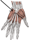
-boxer’s frx (pic): frx of 5th MC w/ volar (palm of hand) displacement of MC head, due to striking an object w/ a clenched fist

Dupuytren’s contracture
cause: progressive thickening/shortening of palmar fascia, nodules develop in palmar aponeurosis, limiting finger extension, ultimately causing flexion deformity
S&Ss:often develops in 4/5th finger, flexion deformity at MCPJs and Prox. IPJs
management: surgical excision of fibrotic tiss
tenosynovitis
pain, swelling, difficulty in movement, spread of infection from 1st and 5th flexor sheaths into the forearm
inflammation of synovial sheath

ulnar nerve injury at wrist
causes: compression between pisiform and hamate - handlebar neuropathy, penetrating wound, injury posterior of medial epicondyle
sensory loss: hypoesthesia in med. 1.5 digits
motor loss: weakness of intrinsic muscles ulnar nerve supplies
deformity: prominent ulnar claw
- 4th and 5th MCPs hyperextended (due to unopposed ex. dig.)
- 4th and 5th IPs flexed (unopposed FDS and FDP)
- flat hypothenar eminence
- > however, in ulnar paradox, the ulnar nerve is lacerated further up the arm beyond the point where it innervates the FDP -> the med. 2 digits are extended and look better when in fact the injury is worse
Treatment: padding (gloves), ice, NSAIDs
articulations and ligaments of elbow joint
Articulations: ant: head of radius w/ capitulum of humerus, trochlea of humerus w/ trochlear notch of ulnar
post.: olecranon process of ulna w/ olecranon fossa of humerus
Ligaments: 2 radial ligaments, 1 ulnar ligament - anterior, posterior, oblique, annular ligament around head of radius, all stabilise
pulled elbow
in children esp girls, eg when pulled up a kerb
weak annular ligament and under development of head of radius, forearm is abducted and med. rotated
subluxation of joint
gentle supination and compression needed and, after a few days, the radius pops back in
4 frxs to humerus and nerves damaged
frx to surgical, anatomical neck of humerus, axillary
mid humeral frx, radial
supracondylar frx, median
ulnar nerv is also vulnerable as it lies unprotected behind the medial epicondyle
nerve routes passing distal humerus
radial: anterior to lat. epicondyle
median: anterior to med. epicondyle
ulnar: posterior to med. epicondyle
medial and lateral epicondylitis
medial/golfer’s elbow: repeated use of flexors, pulls on periosteum of med. epicondyle
lateral/tennis elbow: repetitive use of superficial extensors of forearm, pain radiates down post. apsect of forearm as it can lead ot radial nerve compression
-inflammation of periosteum of lateral epicondyle (ie the epicondylitis)
student’s elbow
repeated pressure on and inflammation of olecranon bursa, local anaesthetics, drainage, steroids, surgical excision
subtendinous bursitis
not common, deeper bursa, triceps tendon and olecranon involced, can lead to tendon calcification, deeper bursa than one in student’s elbow
olecranon frx
fall on elbow, triceps pulls olecranon prximally, surgical intervention - pin, ulnar nerve can be damaged
ligaments of shoulder joint
gleno humeral ligaments reinforce anterior aspect of capsule, coracohumeral and coracoacromial strengthen superiorly
shoulder dislocation
normally anterior, fall on outstretched hand
painful arc syndrome
supraspinatus tendinitis, subacromial bursitis
on ACTIVE abduction, scapulo-humeral rhythm is disturbed
pain is worsened on abduction between ~60-120 degrees, either side of this pain is not there
origins and insertions of rotator cuff muscles
Supraspinatus: supraspinous fossa of scapula -> superior facet of greater tubercle of humerus
Infraspinatus: infraspinous fossa of scapula -> middle facet of greater tubercle
Teres minor: middle part of lat. border of scapula -> inferior facet of greater tubercle
Subscapularis: subscapular fossa -> lesser tubercle of humerus
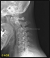
whiplash associated disorder/WAD
h.flexion -> h.extension -> sp/train cervical tissues, can occur when going slowly - 10mph, 1-4 seriousness (4 most), cervical spine most at risk from sideways impact (less ligamentous support), cervical lateral x ray - loss of lordosis due to neck muscles spasming, may have a lack of symptoms for a few weeks
ligaments of the spine
Anterior Longitudinal ligament: stronger than PLL, gets broader from cervical -> lumbar spine, attaches to periosteum of vert. bodies and does not attach to intervert. discs
Posterior Longitudinal Iigament: weaker than ALL, narrower from cervical -> lumbar spine, serrated margins which attach to the intervert. discs but are free over the vert. bodies, separated from vert. bodies by basivertebral veins

differences between vertebral bodies: atlas and axis
atlas/C1: no body or spinous process, large lateral masses w/ widest transverse processes of cervical vertebrae to hold up the cranium
axis/C2: strongest of cervical vert., two large superior articular facets that the atlas can rotate on, dens/odontoid process projuects superiorly, held in place against post. aspect of ant. arch of the atlas by transverse ligament of the atlas

differences between cervical, thoracic and lumbar:
cervical: small oval body, large triangular vertebral foramen, transverse process had transverse foramina containing vertebral artery and vein, short and bifid spinous process (C3-6)
Thoracic: heart shaped body w/ 1/2 costal facets for articulation w/ ribs, circular and smaller vertebral foramen than cervical, long and strong transverse process w/ a decreasing length between T1-12, long spinous process that slopes postero-inferiorly
Lumbar: massive kidney shaped body, triangular foramen that is larger than thoracic’s but smaller than cervical’s, long and slender transverse process, short, thick and sturdy spinous process
OVERALL: as you go down the vertebral column, the foramen gets smaller, the body gets bigger

kyphosis and lordosis commonly due to
K: OP (anterior wedging of thoracic vertebrae)
L: pregnancy, obesity, or weak abdominal muscles
OA (of spine in particular)
degeneration (wear and tear) or articular cartilage and underlying bone, often affects unilateral joints, aggravated by movement, relieved by rest
-xray findings: joint space narrowing, subchondral sclerosis, osteophytes, subarticular cysts, bone spurs in spine
commonest form of arthritis, risk factors include age, obesity, previous trauma
mainly affects knee, hand, hip and pine joints (whereas RA is mainly hands

RA (of spine in particular)
autoimmune, systemic inflammatory disease, affects large joint bilaterally, atlanto-axial instability, commonly affects hand joints
x ray findings: decreased joint space (like OA), marginal erosions (different to OA), osteopenia (low bone mineral density), joint subluxation, soft tissue swelling
in picture the axis and atlas have sublaxed

scoliosis
lateral deviation
structural causes: fixed, fails to correct, can be due to hemivertebra (vertebra not completely developed on one side, wedge shaped), OP, developmental problems
non structural causes: can be due to leg length inequality, weak muscles

ankylosing spondylitis
fusion of joints, similar to RA in that it affects 2 joints and gets better after exercise and worse after rest
a systemic inflammatory disease most likely in young males, linked w/ UC and chron’s (HLA-B27 antigen)
when severe it can lead to fusion of facet joints, SI (sacroiliac) joints
tiredness, malaise, initial weight loss
tests: blood CRP, HLA-B27, MRI

herniation of IV discs
nucleus pulposus ruptures through the annulus fibrosis (jam doughnut analogy), most commonly at L5/S1, then L4/5, commonly leads to flexion
compression of ventral root -> motor symptoms
classic sciatica symptoms (compression of spinal roots that lead to sciatic nerve), ventral and dorsal horn affected
assess the neurological function of limbs (sensory, motor, reflex), SLR (straight leg raise), palpate spine
SLR: lie on back, relax leg in hand, raise leg, pain above 30 degrees

cauda equina
medical emergency, need urgent decompressive surgergy
centro-posterior herniation of IV discs or spinal tumour
symptoms: saddle anaesthesia (numbness around genitalia and buttocks), urinary a/o faecal incompetence (due loss of anal tone, though it may just be changing bowel habit), neurological deficit - functional abnormality of a body area due to a decrease in the function of the brain, spinal cord, muscles or nerves (normally bilateral)
relationship between spinal nerves and vertebra
there are 7 cervical vertebrae, but 8 cervical nerves -> the nerves are always located on top of the vertebra
nerve C8 lies between vertebrae C7 and T1
after that, the nerve lies underneath its vertebra
roles of the ligaments of the hip joint
iliofemoral: ant. and sup., prevents h.extension
pubofemoral: ant. and inf., prevents over abduction
ischiofemoral: post., weakest and has little function
ligament of the head of the femur: from acetabular notch -> fovea for the ligament of the head of the femur, little function in strengthening joint, but contains the artery to the head of the femur, which supplies the epiphysis especially in children
femoral canal
borders: anterosuperiorly inguinal ligament, post. pectineal ligament lying anterior to the superior pubic ramus, med. lacunar ligament, lat. femoral vein
canal has space for the femoral vein to dilate during exercise, also contains lymphatic vessels and lymph nodes embedded in areolar tissue
mid inguinal point/midpoint of inguinal ligament
midpoint directly between ASIS and pubic symphysis, femoral pulse found here/midpoint along the inguinal ligament length, used for a femoral nerve block (injection of local anaesthetic for pain relief)
types of hip frx
subcapital: intracapsular, high up neck, near head, can tear and damage joint capsule and disrupt blood supply
cervical: neck, w/in margins of capsule
- in either of these, retinacular arteries from medial circumflex artery disrupted -> blood supply halted -> avascular necrosis as only artery to head of femur remains
- >commonest in elderly, injured limb appears shorter and laterally rotated (frx large injury -> lat. rotated, disloc. medium injury -> med. rotated)
pertrochanteric: extracapsular, intertrochanteric region
dislocations of hip
acquired: posterio-inferior dislocation - when hip is flexed + adducted + medially rotated
- presentation is limb shortening and med. rotation
- can cause compression of sciatic nerve leading to paralysis of hamstring muscles and muscles distal to knee, sensory deficits in leg
congenital: due to abnormal angle of neck of femur, most common in girls
+ve trendelenburg
caused by superior gluteal nerve lesion, frx of greater trochanter (glut med attaches here), dislocation of hip joint
abductors maintain a level position when opposite limb is unsupported (in swing phase), results in waddling gait (raising pelvis), steppage gait (lifting foot) or swing-out gait (foot out laterally)
possible causes of swelling in popliteal fossa
popliteal aneurysm (pulsatile swelling)
baker’s cyst (swelling of semimembranosus tendon or herniation of synovial membrane)
abscess
tumour
enlarged lymph nodes
unhappy triad
lateral impact during extension
medial meniscus, medial collateral and anterior cruciate damaged
can tell by performing ant/post draw test, collaterals
menisci tears
in young people it can be due to a sporting injury, twist of knee/direct tackle, in the elderly it is weakened w/ age
True locking: something physically gets in the way stopping extension eg meniscus, stuck in about 20-30 degrees of flexion, can flex beyond that, but extension is stopped
Pseudo locking: pain inhibits knee movement, can be due to inflam arthritis, gout, septic arthritis
dislocation of patella
direct blow/sudden twist of knee
weakness invastus medialis causes the patella to dislocate laterally w/ a lot of pain and effusion (seeping of fluid into body cavity)
more common in women due to an increased q angle (angle between line from ASIS -> patella and patella -> tibial tuberosity, in women it should be less than 22 degrees, in men less than 18)
pulled hamstring
common in those who run a/o kick hard eg running, jumping, football
violent muscular exertion can tear part of the prox. attachments to the ischial tuberosity
extremely painful
bursa/bursitis of knee
pre-patellar bursa -> housemaid’s knee (caused by one off large injury eg fall onto knee, or many minor injuries commonly from kneeling)
subcutaneous infrapatellar bursa -> clergyman’s knee (from kneeling a lot again, esp. on hard surfaces which is what clergymen used to do)
suprapatella bursa
deep infrapatellar bursa

compartment syndrome
raised intra-compartmental pressure -> intra arterial pressure -> arterial occlusion -> ischaemia
presentation: 6 Ps: pain, paresthesia, perishing cold, pallor, pulselessness, paralysis
roots of sciatic nerve, supply and termination
sacral plexus (anterior and post. divisions of ant. rami of L4-S3)
supplies all hamstrings and hamstring portion of adductor magnus
-also indirectly lower leg and foot sensory innervation
bifurcates into tibial (all hamstrings) and common fibular (short head of biceps femoris)
roots of gluteal nerves, supply and termination
Superior: post. L4-S1, glut med, min and TFL, enters sup. to piriformis and goes as far as TFL
Inferior: post L5-S2, glut max, divides into several branches at the inf. part of glut. max.
pott’s frx
mech. forced foot eversion
deltoid ligament pulls on med. mall. and causes avulsion
talus displaced lat. and post. and causes avulsion of lat. mall., bimalleolar frx
varicose veins
long dilated tortuous superficial veins due to incompetent valves at sapheno-femoral/-popliteal or perforator junctions (where perforator veins enter muscles)
-venous stasis/stagnation is a cause of this, and can be due to a) incompetent/loose fascia that fails to resist muscle expansion, b) external pressure eg bedding in hospital or a tight cast or c) muscular inactivity eg from a long flight
risk factors: prolonged standing, obesity, pregnancy, family history, oral cont. pill
surgery can be used
medial malleolus relations to vessels
posterior: Tom (tibialis posterior), Dick (FDL) and (post. tibial artery) very/bloody (posterior tibial vein) nervous (tibial nerve) Harry (FHL)
anterior: long saphenous vein, saphenous nerve
apparent and true limb shortening
Apparent: xiphisternum/xiphoid process or umbilicus-> med. malleolus
-looks at eg fixed joint deformity, pelvic tilt
True: from greater trochanter of femur/ or ASIS -> med. malleolus
-looks at actual loss of bone length
why does a peripheral nerve lesion -> skin becoming red -> dry and scaly?
peripheral contains sensory, efferent and autonomic fibres, loss to SyNS fibres can lead to loss of vasoconstriction to skin -> vasodilation and redness from increased blood flow to the skin
-> loss of innervation to sweat glands -> dry then scaly
flexor retinaculum attachments
medially: pisiform and hook of hamate
laterally: tubercle of scaphoid and med. part of volar surface and ridge of trapezium
femoral ring borders and contents
lat. : vert. septum between femoral canal and vein
post. : superior ramus of pubis covered by pectineus muscle and fascia
med. : lacunar ligament
ant. : med. part of inguinal ligament
contents: base of the femoral canal