Revision: vasculature Flashcards
(37 cards)
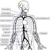
name major arteries


name major veins


name major coronary arteries and cardiac veins

anterior interventricular=left anterior descending

name major coronary arteries and cardiac veins of posterior heart


Describe the vasculature of the U/limb from the subclavian arteries
Subclavian -> Axillary artery at the lateral border of the 1st rib
Axillary becomes the posterior and anterior humral circumflex arteries that supplies the shoulder, as well as the subscapular artery, all at the level of the humeral surgical neck
Axillary becomes the brachial at the lower border of the teres M.
Brachial provides the profunda brachii immediately after the teres M that runs down the radial groove and ends by contributing to a network of vessels at the elbow
After the cubital fossa the brachial artery divides to the ulnar and radial arteries that supply the ant. and post. of the forearm, respectively
these then both form their own superficial and deep palmar arches that anastamose together- the superficial arch produces the common palmar digital arteries, that then divide to form the proper palmar digital arteries
desc. clinical significance of arteries of the u/limb
Axillary aneurysm:
- caused by high BP, Marfan’s, baseball pitchers
- puts pressure on the brachial plexus, this then causes pain and anaesthesia in the cutaneous distribution of the affected nerve: axillary - regimental badge area, radial - back of arm and forearm as well as lateral dorsum of hand, ulnar: medial palmar and dorsum of hand, median: lateral palmar hand, muscolcutaneous: lateral forearm
Occlusion/laceration of brachial artery:
- this is an emergency as it can lead to infarction, paralysis, muscles being replaced by scar tissue and shortening leading to flexion deformity, Volkmann’s contracture:
- > permanent flexion contracture of hand at wrist, passive extension of fingers is painful and restricted, hand is blue/white and cold, radial pulse is absent, claw-like deformity of hand and fingers
name vessels


name vessels


name vessels


name vessels

proper palmar arches are at the top

desc. veins of U/limb from cubital and basilic to axillary and clin. significance
cephalic runs down antero-lateral side of U/limb, basilic runs more medially
both are superficial and can normally be seen, connected by median cubital vein, used for venepuncture as it is relatively superficial and easily accessible
others run alongside the major arteries -> the vena comitantes
The basilic becomes the axillary vein at the lower border of the teres M, the cephalic joins the axillary between the pec M and deltoid
name vessels


name vessels


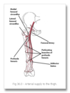
desc. arteries of L/limb
Common iliac -> external and internal iliac:
internal -> (branch) obturator -> pelvic region via obturator foramen -> (medial thigh) post branch (distal deep gluteal muscles) and ant. branch (pectineus, obt. ext., adductors, gracillis)
external -> femoral as it passes the inguinal ligament and enters fem. triangle
fem. artery produces a branch called the profunda femoris -> perforating branches (3/4 arteries that perforate the add. mag. and supply some muscles in the medial and posterior thigh), lat. fem. circumflex (wraps around ant. femur and supplies some muscles in lat. side of thigh) and med. fem. circumflex (wraps around post. femur and supplies head and neck)
- the lateral circumflex supplies less blood to the femoral head as it has to penetrate the iliofemoral ligament
femoral artery moves down the ant. side of the thigh down the adductor canal (supplying add. muscles) and becomes popliteal artery as it moves through the adductor hiatus
popliteal artery gives off genicular branches that supply the knee joint and splits to form posterior and anterior tibial arteries
post. tib. artery: gives off fibular artery that supplies lat. side of leg, the rest of the post. tib. artery supplies the post. side of the leg
anterior tibial: supplies all muscles of ant. leg, moves through IO membrane and becomes the dorsalis pedis as it passes the ant. aspect of the ankle joint
desc. clinical significance of arteries of L/limb
med. fem. circumflex artery wraps round the post. side of the femur and supplies the head and neck, in a fem. head frx this can be damaged causing avasc. necrosis of femur head
Athrosclerosis-> intermittent claudication
-Pain on walking that is then relieved by rest, a build up of an atherosclerotic plaque (therefore same risk factors as for CAD), limits flow to muscles -> ischaemia and pain
Pulses: easiest is femoral artery, midway between ASIS and pubic symphysis (ie. in the inguinal ligament)
popliteal is hardest as it is deep within the popliteal fossa, an aneurysm here forms an obviously palpatable pulsation in the popliteal fossa, which can lead to damage to the tibial nerve -> motor loss of post. leg muscles and anaesthesia
the dorsalis pedis can be palpated on the dorsum of the foot, medial to the EPL

name vessels


name vessels


name vessels


desc. veins of the L/limb from dorsal venous arch to external iliac
The dorsal venous arch mainly drains into superficial veins, the most important two being the Great and Small Saphenous veins, but it also forms deep veins, the anterior tibial:
GSV: from med. part of Dorsal venous arch, moves along medial leg anterior to the medial malleolus and post. to med. condyle in knee, drains into femoral vein just distal to the inguinal ligament
SSV: from lat. part of dorsal venous arch, moves up the post. leg, post. to lat. mall. and along lat. border of calc. tendon, it moves between the lat. and med. heads of the gastrocnemius and drains into the popliteal vein in the popliteal fossa
Deep veins: the anterior tibial vein comes from the dorsal venous arch, along with the post. tibial and the fibular vein it forms the popliteal vein that then forms the femoral vein as it passes through the adductor hiatus
The deep vein of the thigh drains into the femoral vein
The femoral vein becomes the external iliac vein at the level of the inguinal ligament
name vessels

long = great saphenous veins

name vessels


name vessels


name vessels


clinical significance of veins of l/limb
Varicose veins: incompetent valves lead to superficial vein dilation
- this causes inc. pressure in veins -> damages cells, blood exudates to skin
- further problems can lead to brown pigmentation and ulceration
- treatments include: surgical movement of saph. system, reconstruction of valves, tying off affected valves
DVT: formation of blood clot in deep veins of l/limb, can lead to pulm. occlusion from the thrombus embolising and reaching the pulm. circulation, leads to cardio-pulmonary arrest
- can also lead to postphlebitic/post-thrombotic syndrome->chronic deep venous insufficiency, damage to venous valves, lymphoedema
- Prophylaxis: leg compression given in surgery, subcutaneous heparin
- Treatment: IV heparin, oral warfarin
Great saphenous vein can be used for venepuncture, skin incision anterior to medial malleolus, risk of saphenous nerve injury and pain along medial border of foot


