Respiratory Fungi Flashcards
Systemic Mycoses
Histoplasma, Coccidiodes, Blastomyces, Paracoccidioides
- Primary pathogens
- Severe disease in immunocompromised hosts
-
Airborne/Environmental transmission
- No person-to-person transmission
- Starts in the lungs but frequently disseminates to viscera
- Endemic to certain geographic areas
-
Dimorphic
- Molds @ 25°C
- Yeast or spherules @ 37°C
Histoplasma capsulatum
Characteristics
- Causes Histoplasmosis
-
Dimorphic
-
Mold grows in soil w/ high N2 content
- Bat and bird droppings promote growth
- Filamentous septate hyphae
- Produces spores ⇒ micro and macroconidia
- Macroconidia = tuberculate chlamydospores
- Yeast grows @ 37°C within Mφ
-
Mold grows in soil w/ high N2 content

Histoplasma
Transmission
Inhalation of microconidia
(Asexual spores)
Causes histoplasmosis.

Histoplasmosis
Epidemiology
Ohio and Mississippi Valleys
&
Central and South America
Outbreaks associated with bird roosts, caves, urban renal projects involving excavation.

Histoplasmosis
Clinical Presentation
- Asymptomatic infection ⇒ 90%
-
Flu-like sx ⇒ 5%
- Fever, chills, malaise, HA, lymphadenopathy
- Some progress to pulmonary histoplasmosis ⇒ 1%
- Cough, SOB
- Resembles TB w/ caseating granulomas
-
Progressive disseminated disease ⇒ 1%
- Immunocompromised hosts
- Found in 10-25% of AIDS pts living in endemic areas

Histoplasmosis
Pathogenesis
Disease manifestations depend on innoculum size & host immune status:
- Microconidia enter lung
- Converts to yeast form
- Phagocytosis by alveolar MΦ
- Replicate within MΦ
- Facultative intracellular parasites
- MΦ transport yeast to regional LN
-
Transient fungemia
- Skin, liver, spleen common sites
- Does not necessarily mean disseminated disease
- Can be seen even with mild cases
-
T-cell sensitization ⇒ fungicidal activity of Tc & MΦ
- Can take 6-8 weeks
- Subsequent granuloma formation
- Clinical picture frequently resembles TB
- Resolution or progression
- Possible reactivation when immunity wanes

Histoplasmosis
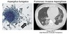
Diagnosis

Histoplasmosis
Treatment
-
Mild pulmonary disease
- Self-limiting
- No treatment
-
Severe acute pulmonary disease
- Itraconazole
-
Systemic disease
- Amphotericin B followed by oral itraconazole
Histoplasma vs Tuberculosis
- Both histoplasmosis & TB
- Primary pathogen w/ severe disease in immunocompromised
- Facultative intracellular organisms
- Infection by respiratory route
- Pulmonary infection that can disseminate
- Can detect infection by skin testing
- Reactivation can occur
- Mtb only ⇒ person to person transfer
Coccidioides immitis
Characteristics
- Causes Coccidiomycosis
- AKA Valley Fever or Desert Rheumatism
- Most virulent of the mycoses
-
Dimorphic
-
Arthrospores grow in warm, alkaline soil
- Barrel-shaped septate hyphae
- Growth enhanced by bird droppings
-
Spherules containing endospores grow @ 37°C
- Extracellular pathogens
-
Arthrospores grow in warm, alkaline soil

Coccidioides
Epidemiology
- Prefers soil of arid regions
- San Joaquin Valley
- Southwest US
- Mexico and parts of Central and South America
- Conditions that favor aerosols promote outbreaks
- Cycles of heavy rain followed by draught
- Warming trends leading to dispersal
- 150k new infections/yr
- 80% of longterm residents infected
- Non-immune visitors/new residents at risk

Coccidioides
Transmission
Inhalation of arthrospores (arthroconidia)

Coccidioides
Risk Factors
High risk for chronic/disseminated disease:
- > 65 y/o
- HIV
- 1st trimester of pregnancy
- DM
- Native Americans, Filipinos, African Americans, Hispanics
Coccidioides
Pathogenesis
- Most virulent of systemic mycosis ⇒ only need a few for infection
- Arthrospores (conidia) inhaled into mid-lung area
- Temperature causes conversion into a large spherule w/ many small endospores
- Extracellular pathogen
- Spherule rupture releasing endospores ⇒ ± dissemination
- Requires cell-mediated immunity to eradicate spherules

Coccidioides
Clinical Presentation
- Asymptomatic ⇒ 50-60%
- Flu-like illness ⇒ called Desert Rheumatism, Valley Fever, or Primary coccidiomycosis
- Cough, CP, weight loss, arthralgia, skin rash
- Can last for weeks to months
-
Severe/chronic pulmonary disease ⇒ 5-10%
- Cavitary disease common
-
Disseminated disease ⇒ 1%
- Also called chronic meningitis
- Fatal if untreated
- Requires lifelong treatment
Coccidioides
Diagnosis
-
Skin test ⇒ reactivity within 2 weeks
- Anergy in disseminated disease
- Indicates infection has occurred but not neccessarily responsible for present illness
- Biopsy ⇒ spherule forms
-
Culture ⇒ mold forms
- Sputum or tissue sample
- Grown on Sabouraud’s agar
- Takes 2-3 days
- Lab acquired infections a problem
-
Serology
- CSF ⊕ within 2-3 weeks
- Rising titers after 2-3 months suggest dissemination

Coccidioides
Treatment
- Most difficult of systemic mycoses to treat
-
Recurrent/relapse disease is common
- Reinfection after successful treatment
- 12-24 months of azole depending on severity
- Amphotericin B for severe disease
- Chronic meningitis ⇒ amphotericin B then switch to azole for life
Blastomyces dermatitidis
Characteristics
- Causes Blastomycosis
- Morphology ⇒ dimorphic
- Septate hyphae and asexual spores @ 25°C
- Broad-based budding yeast cells @ 37°C

Blastomyces
Epidemiology
-
Endemic east of the Mississippi River
- Esp. Arkansas, Kentucky, Louisiana, North Carolina, Tennessee, Mississippi
- Spores grown in soil high in decaying organic matter
- E.g. wooded areas
- Outbreaks via occupational or recreational contact w/ soil
- See disease commonly in 20-50 y/o males

Blastomyces
Transmission
Inhalation of asexual spores
Natural disease in dogs and horses.
Blastomyces
Pathogenesis
- Inhaled conidia (asexual spores)
- Yeast forms in lung within MΦ
- See a mixed granulomatous & pyogenic response (abscess)
- Similar to TB & bacterial PNA
-
Dissemination may involve skin and bone but is rare
- Not a common opportunist in immunocompromised but causes severe disease when it occurs
- Cell-mediated immunity critical but PMNs also play a role in clearing infection
- Reactivation possible

Blastomyces
Clinical Presentation
- Asymptomatic to flu-like illness
-
Pulmonary form
- Similar to bacterial PNA ⇒ infiltrates but no calcification
- Cough and CP
-
Dissemination with or without pulmonary resolution
- Skin, joints, bone, CNS
- Ulcerative lesions of skin and bones

Blastomyces
Diagnosis
- ID organism in tissue
- Culture - takes > 1 week
- Mold form
- Yeast form (broad-based budding yeast)
- Ag based tests

Blastomyces
Treatment
Treatment depends on severity of infection and host immune status.
Moderate disease ⇒ Azoles (Ketoconazole, itraconazole)
Disseminated disease ⇒ Amphotericin B