COPD Flashcards
COPD
Definition
Disease state characterized by airflow obstruction that is not fully reversible.
Usually progressive and associated with an abnormal inflammatory response of the lungs.
Umbrella term for many different disease processes:
- Chronic bronchitis
- Emphysema
-
Asthma
- ~ 15% of COPD patients have a reactive airways component ⇒ “asthma-COPD overlap syndrome”
- Bronchiectasis
- Cystic fibrosis
COPD
Epidemiology
- Most common pulmonary disease ⇒ 32 million in the US
- Leading cause of morbidity and mortality worldwide
-
3rd leading cause of death in the US
- ~ 20,000 deaths/yr
- Greatest cause of missed days of work
- 2nd highest cause of disability
-
3:1 male to female ratio
- Women morely likely to have COPD in countries with higher socioeconomic status
COPD
Etiology & Risk Factors
-
Tobacco smoke ⇒ leading cause of COPD
- 15-20% of long-term smokers develop COPD
- Exposure to secondhand smoke
- Inhalation of smoke ⇒ from biomass fuel or ambient particle matter
- Indoor air pollution ⇒ from heating and cooking with biomass in poorly ventilated dwellings
- Outdoor air pollution
- Alpha-1 Anti-trypsin deficiency
- Occupational dusts and chemicals
- Infections ⇒ especially severe childhood respiratory infections
- Airway hyperresponsiveness
- Gender
- Age
- Socioeconomic status
Smoking Effects
Smoke inhalation leads to:
- ⊗ Ciliary mobility
- ↑ Airway resistance by reflex bronchoconstriction
- Altered MΦ function
- Squamous metaplasia
- Mucus gland hyperplasia

COPD
Pathogenesis
Not fully understood but multifactorial.
-
Hypersecretion of mucus
- Likely a response to irritation
- Impairment of lung clearance
- Destruction of bronchial tissue
- Alterations of lung defenses
- Primary degenerative changes in collagen and elastic tissue of alveolar septa (emphysema)
-
Lung elastin damage by elastase
- Normally present in lungs but also secreted by MΦ and PMNs
- Countered by elastase inhibitors in serum (alpha-1-antitrypsin)
- Imbalance or malfunction ⇒ excess elastase ⇒ destruction of elastin ⇒ loss of elastic recoil
COPD
Pathophysiology
Airflow obstruction results from:
-
Small airways disease
- Airway inflammation
- Mucus gland hyperplasia
- Fibrosis, airway narrowing
- Loss of airway tethering
-
Lung parenchymal destruction
- Dilation/destruction of bronchioles
- Enlargement and/or destruction of acinus
- Decreased elastic recoil
Chronic Bronchitis
Definition
Clinically defined disease with increased mucous production and chronic or recurrent productive cough.
Cough:
Frequency ⇒ > 3 months per year
Duration ⇒ > 2 years

Emphysema
Definition
Morphologically defined disease with enlargement of air spaces due to destruction of alveolar septae.
⇒ Leads to loss of airway structural supoort resulting in functional obstruction to air flow
⇒ Different morphologic types

Chronic Bronchitis
vs
Emphysema

Chronic Bronchitis and Emphysema
Pathogenesis

Emphysema
Pathogenesis
Permanent and abnormal enlargement of terminal respiratory unit of acinus.
- Described as “lung dry rot”
- Alveolar distention
- Destruction of alveolar septae
- Tobacco use produces ROS that can inactivate antiproteases ⇒ “functional” α1AT deficiency

Emphysema
Types
Two main types:
- Centriacinar (aka Centrilobular - CLE)
- Panlobular (PLE)

Centriacinar
Emphysema
“Centrilobular Emphysema (CLE)”
- Selective dilatation of respiratory bronchioles and alveolar ducts
- Most common type
- Usually smoking related
- Most severe in upper lobes
- See enlarged air spaces of varying sizes

Panlobular Emphysema
(PLE)
- Dilatation of all components beyond the terminal bronchiole
- Usually occurs in the lower lung
- Severe forms in young people usually associated with α1AT deficiency
- See enlarged airspaces of uniform size

CLE vs PLE

Bullous Disease
“Paraseptal Emphysema”
- See > 1 cm dilatations of air spaces in the periphery of the lung adjacent to pleura
- Occurs adjacent to scars with aging or from unknown factors
- Does not qualify as true emphysema

Emphysema
Alveolar Changes

Emphysema
Expiration Changes

Chronic Bronchitis
Pathogenesis
May have no demonstrable pathology
or
May have any of the following:
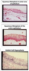
- Smoking related changes
- Squamous metaplasia
- Loss of cilia in bronchial epithelium
-
Hyperplastic bronchial mucus glands with chronic excessive mucus secretion
- Measured using Reid index
- Inflammatory infiltrates of bronchial wall
- Globlet cell hyperplasia
- Fibrosis and atrophy of bronchial walls
- Smooth muscle hyperplasia
- Bronchial hyperresponsiveness (50% of COPD)

Chronic Bronchitis
Smoking Effects
Occurs in cigarette smokers 10-12 years after starting.
Affects 10-15% of smokers.
Reid Index
Ratio of bronchial glands to the entire thickness of bronchial wall from epithelium to top of the cartilage.
Method for measuring degree of mucus gland hyperplasia.

Chronic Bronchitis
Histology

Chronic Bronchitis
Secondary Infections
- Infections are common in the bronchi
- Leads to further destruction of bronchial tissue
-
Haemophilus influenzae colonizes airway of smokers
- Creates an immune reaction in airways
- Isolated from sputum in 60% of stable chronic bronchitis pts
COPD
Symptoms
Ranges in severity and may be applicable to all disease types.
- Cough ± sputum production
- Dyspnea ⇒ initially on exertion, later at rest
- Wheezing
- Decreased exercise tolerance
- Weight loss and lack of appetite ⇒ late
- Impaired mentation and fatigability ⇒ late and usually when cor pulmonale has occured
COPD
Physical Exam
- Slightly prolonged forced expiration
- Decreased breath sounds at apices or bases
- Scattered expiratory rhonchi and wheezes
- Use of accessory muscles
- Hyperresonance
- ↑ AP diameter of chest
- Low diaphragm
- Inaudible heart sounds
- Signs of cor pulmonale
- Heart failure with JVD
- Functional tricuspid insufficiency
- Hepatojugular reflex
- Hepatomegaly
COPD
Radiographic Changes
Often normal but may show:
Emphysema ⇒ hyperinflation and/or loss of peripheral markings
Chronic bronchitis ⇒ increased bronchial markings

Blue Bloater
Seen with chronic bronchitis

Stocky and obese
Markedly hypoxic and cyanotic
Pink Puffer
Seen with emphysema:

Tachypnea
Prolonged expiration and pursed lip breathing
Hyperresonant chest
Diminished breath sounds
Cachexia
Chronic Bronchitis vs Emphysema
Summary

COPD
Diagnosis
- Consider COPD if patient has symptoms of:
- Dyspnea
- Chronic cough or sputum production
- Risk factors for the disease
-
Spirometry required for diagnosis
- FEV1/FVC (FEV-1%) < 0.70 and FEV1 ≤ 80%
COPD vs Asthma
Diagnosis

COPD
Disease Staging
In patients with FEV-1%:
- GOLD 1: Mild ⇒ FEV1 ≥ 80% predicted
- GOLD 2: Moderate ⇒ FEV1 between 50-80% predicted
- GOLD 3: Severe ⇒ FEV1 between 30-50% predicted
- GOLD 4: Very severe ⇒ FEV1 ≤ 30% predicted
COPD
Comorbidities
COPD patients are at increased risk for:
- Cardiovascular disease
- Respiratory infections
- Osteoporsis
- Anxiety and depression
- Diabetes
- Lung cancer
- Bronchiectasis
COPD Treatment
Preventative Measures
- Smoking cessation ⇒ can greatly affect outcomes
- Reduce indoor air pollution ⇒ use stove instead of open fire
-
Influenza and pneumococcal vaccination
- Recommended for COPD patients
- May reduce exacerbations or hospitalizations
-
Pulmonary rehabilitation
- Reduces hospitalizations and mortality in recent COPD exacerbations
-
Physical activity
- Possibly as effective as smoking cessation
- Physical inactivity is likely very detrimental
COPD Treatment
Maintenance Pharmacotherapy
Effectiveness
- Improves quality of life
- Improves COPD symptoms
- Reduces exacerbations
- Improves exercise tolerance
- Does not improve lung function decline long-term
Bronchodilators
Overview
- Reduces exacerbations and hospitalizations
- Improves symptoms and health status
- Combining different classes may improve efficacy
- Inhaled are best and safest
- Long-acting more convenient and effective
Bronchodilator
Classes
-
β-agonists
-
SABA ⇒ short-acting
- Albuterol
-
LABA ⇒ long-acting
- Salmeterol
- Formoterol
-
SABA ⇒ short-acting
-
Anti-cholinergics (muscarinic antagonists)
-
SAMA ⇒ short-acting
- Ipratropium
-
LAMA ⇒ long-acting
- Tiotropium ⇒ appears superior to LABA in recurrent exacerbations
-
SAMA ⇒ short-acting
Inhaled Corticosteroids
- Does not appear to slow decline in lung function
- May decrease frequency of exacerbations if FEV1 > 60%
- Often used in combo with LABA or LAMA
- Long term use may increase risk for PNA
- Withdrawal from treatment may lead to exacerbations
Combination Therapy
ICS + LABA
or
ICS + LABA + Anticholinergic (tiotropium)
- Appears to provide even more benefit
- ↓ Exacerbations in moderate-severe COPD
- Caution in pts with cardiac disease
- Can exacerbate arrhythmias
Roflumilast
- Phosphodiesterase-4 inhibitor
- Used in pts with severe and very severe COPD
- Reduces COPD exacerbations treated with oral steroids
Theophylline
- Phosphodiesterase inhibitor
- Oral or IV only
- Less effective and less tolerated than bronchodilators
-
Low dose theophylline reduces exacerbations
- Does not improve post-bronchodilator lung function
- Poor risk/benefit ratio
- Drug interactions, toxicity
- Risk of osteoporosis and DM
- Use only in select cases
Oral Steroids
Chronic systemic oral steroids not recommended.
Unfavorable risk-to-benefit ratio.
Only used in very severe cases.
Surgical Interventions
-
Lung volume reduction surgery (LVRS)
- More effective than medical therapy for pts with upper-lobe emphysema and low exercise capacity
- High costs
- For very select pts
-
Lung transplantation ⇒ very severe COPD
- Can improve quality of life and functional capacity
-
Bullectomy ⇒ for giant bullae
- Can improve dyspnea
COPD Treatment
Summary
- Long-acting preferred over short-acting
- ICS + LABA/LAMA recommended for pts at high risk for exacerbations
- Steroid monotherapy (inhaled/oral) not recommended
- Roflumilast may be useful to reduce exacerbations
- Patients with FEV1 < 50%, chronic bronchitis, or frequent exacerbations
- Pulmonary rehab and physical activity encouraged
COPD Exacerbations
- Acute episodes of worsening SOB ± dyspnea, cough, and change in sputum (volume, purulence)
-
Often triggered by infections
- Viral
- Bacterial
- Strep pneumoniae
- Haemophilus influenzae
- Moraxella catarrhalis
COPD Exacerbation
Treatment
-
Mild cases
- Often can be treated at home with rest and inhalers
-
Moderate cases
- Oral prednisone 40mg x 5 days
- Possible abx
- Inhaled bronchodilators
-
Severe cases
- Hospitalization
- IV steroids
- Abx
- Inhaled bronchodilators
- Supportive measures ⇒ O2, NIPPV, intubation
Alpha-1-antitrypsin Deficiency (AATD)
Overview
-
Deficiency of anti-protease alpha-1-antitrypsin
- Allows lung enzymes (elastases) to break down lung proteins
- Creates inflammation ⇒ emphysema
- Accounts for 1% of all COPD cases
-
Autosomal co-dominant transmission
- PI*MM ⇒ normal
- PI*ZZ ⇒ homozygous for z allele
- PI*M or null alleles ⇒ varying risks of COPD
Alpha-1-Antitrypsin Deficiency
Clinical Presentation
Suspect and test for AATD in patients with:
- COPD in a never smoker
- COPD development at < 50 y/o
- Strong Fhx of COPD
- COPD with lung basilar predominance on CXR
- COPD with associated unexplained liver disease
Alpha-1-Antitrypsin Deficiency
Diagnosis
Serum Alpha-1-Antitrypsin level
Genetic testing
Alpha-1-Antitrypsin Deficiency
Treatment
Same as regular COPD + replacement therapy
• Intermittent IV infusions of α1AT appears to slow rate of decline in lung function in pts with airflow obstruction