Renal Anatomy Flashcards
What is the blood supply to the kidneys?
- comes from the abdominal aorta through renal arteries
- drain through renal vein (25% of blood you have right now is going to kidneys so this vein is large)

-all of these structures are retroperitoneal


- gonadal arteries are long because gonads develop high up
- kidneys and gonads pass each other during development when gonads descend and gonads ascend
- right kidney should be lower than left
- bladder sits behind pubic symphysis and starts to come over the top when it fills up


- liver moves a lot when you breathe
- kidney moves in too
- perirenal fat provides some cushioning
- only half of kidney is protected by ribs (if you get blunt force to the back, these could pierce kidney)


What is the renal hilum?
- hilum is area where things enter and exit organ
- area where fat goes into kidney
- where blood supply comes in (arteries in, veins out) and ureter goes out
What are the renal cortex, renal colums, and renal medulla?
Renal cortex:
- glomeruli located here
- create filtrate
Renal medulla:
- columns: extension of renal cortex into medulla
- renal pyramids
- renal papilla is where filtrate ends up and is collected by calices

How do we produce urine?
- comes out of collecting duct of nephron from renal cortex
- collecting duct ends in papilla and drains into minor calyx, major calyx, then renal pelvis
- goes through ureter to the bladder

What is a polycystic kidney?
- instead of being smooth on the surface, it has fluid filled sacs
- needs to be transplanted

- infection can pass easily from one kidney to another
- hydronephrosis: built up fluid, swollen
What are supernumerary renal arteries?
- 25% of major malformations in kids are urogenital
- 2 renal arteries on one side, 3 on the other

What occurs in the renal tubule?
- renal corpuscle is the glomerular capsule and glomerulus
- glomerulus are tufted capillaries on inside
- to feed these, afferent arteriole brings in blood and an efferent arteriole leaving
- efferent arterioles spread through tubules
- proximal convoluted tubule
- loop of Henle which goes into medulla
- distal convoluted tubule (close to glomerulus)
- juxtaglomerular apparatus samples filtrate at distal convoluted tubule and if something is wrong, the amount of filtrate made by glomerulus is changed
- fluid goes out the collecting duct (can change amount of water in urine)
- past collecting duct, it is called urine because nothing else happens to it

How much of the circulation goes to the kidney?
-20-25% of circulation goes directly to the kidney but the kidney is only 0.5-1% of body weight
How much filtrate is made?
- from the 20-25% of blood flow, 180L is made into filtrate however the filtrate has virtually no protein or formed elements of the blood or any large molecules
- in the end, you only make about 1-2L of urine (about 99% of the fluid is reabsorbed)
- although the filtrate has lots of small molecules like glucose, amino acids, and bicarbonate ions, the urine has basically no protein, glucose, or bicarbonate
What is the big picture of renal function?
- afferent arteriole blood goes in to renal corpuscle and through glomerulus
- blood goes out efferent arteriole through the peritubular capillaries
- filtration occurs at renal corpuscle (small things will get into filtrate, large things stuck in blood)
- reabsorption of material from the filtrate in the tubule to the blood
- in the renal tubule:
- reabsorption of material from the filtrate in the tubule to the blood
- secretion of material from peritubular capillaries into the filtrate

What are the cell types in the juxtaglomerular apparatus?
- macula densa: part of distal convoluted tubule, “tasting” filtrate, make NO
- granular cells: make renin, have granules, if something is detected as wrong by the macula densa the amount of renin can be altered
- mesangial cells: some in JG apparatus and some in between the tufts of capillaries, important in regulating filtration, modified smooth muscle cells by contracting or relaxing


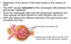
How does blood flow through the renal corpuscle?
- notice that the glomerular capsule is a continuous layer of tissue with the glomerular capillaries jammed into the centre
- the space between the visceral and parietal layer is the capsular space and that is where the filtrate is formed
- with each heart beat, blood is getting pumped through the glomerular capillaries which pushes filtrate out

What structure is on a renal capillary? What is the structure of a renal capillary?
- fenestrated endothelium
- basal lamina (negatively charged from loose aggregations of collagen to act like a -ve charge filter to keep -vely charged proteins in the vascular space)
- podocyte
- has pedicels
- in between the pedicels of the podocytes are filtration slits
- filtrate must move from inside capillary through the fenestrae and filtration slits into the capsular space

Describe the filtration process
- blood flowing through filtration slits into capsular space
- all small molecules can pass through capillary to capsular space
- large molecules ( > albumin size) cannot and stay in the blood
- as blood flows, 15-20% of plasma becomes the filtrate and the rest goes on to peritubular capillaries


The glomerular capillaries are involved in _____ while the peritubular capillaries are involved in _______.
filtration
absorption and secretion
How do the kidneys and CV system work together?
- the CV system generates the pressure necessary for glomerular filtration and drives the high flow needed to maintain a stable cortical interstitial solute composition
- the kidneys maintain blood volume, regulate plasma osmolality, and secrete mediators that affect both cardiac performance and vascular tone
Describe net filtration pressure
- pressure pushing fluid out of the glomerular capillaries (positive pressures):
- blood, hydrostatic pressure in capillaries (ie. the pressure of the blood in the glomerular capillaries)
- pressure holding fluid in the glomerular capillaries (negative pressures):
- capsular hydrostatic pressure (ie. the pressure of fluid already in the capsule)
- blood osmotic pressure (ie. the attraction of the dissolved materials in the blood for water)
- there is usually net positive pressure of 10mmHg
- this pressure pushes filtrate into the capsular space and this determines the glomerular filtration rate (GFR)