Cardiovascular Development Flashcards
(30 cards)
What is pulmonary circulation?
- right side
- oxygenation and removal of CO2 and waste products
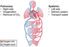
What is systemic circulation?
- left side
- delivery system
- transport oxygen and nutrients, getting rid of waste products

What are key features of post natal circulation?
- two distinct circuits
- oxygen rich is separated from oxygen poor blood
- arranged in series; blood coming from lungs into left side of heart goes into body tissues, oxygen-CO2 exchange at capillaries, return of blood through venous system, goes through great vessels and returns to the right side (output of one side is input of the other)
- pressure is higher on the left side than the right side (systemic>pulmonary)
What are key distinguishing features of fetal circulation?
- mixing of oxygen rich and oxygen poor blood
- 2 open circuits operating in parallel
- gas exchange occurs at the placenta
- pressure on right side > left side (pulmonary>systemic)
- because gas exchange occurs at placenta, lungs don’t need to do work of gas exchange so the lungs are focused more on their developmental role so there is vasoconstriction in pulmonary circulation
- placenta is attached to systemic circulation results in lower pressure on left side (placenta is low resistance vessel)
- pulmonary vasculature is largely closed because lungs are non-functional which causes high pulmonary resistance on the right side


Describe blood flow from the placenta in fetal circulation (bypass)
- umbilical cord to umbilical vein
- ductus venosus is a bypass of the liver; maternal circulation is where filtering of waste happens so fetal liver focuses on development
- inferior vena cava contributes blood from fetal body so this is when mixing happens
- moves into right atrium (also from SVC)
- foramen ovale: opening of septum between left and right side of heart
- blood passes from right side to left side
- preferentially streaming of oxygenated blood to left side of heart
- some blood will also move into right ventricle
- blood from left side also contributed through pulmonary veins
- left atrium to ventricle, aorta, body tissues
- movement of blood from right atrium to right ventricle is normal initially then reaches ductus arteriosus; bypass that connects pulmonary trunk to descending portion of aorta
- lungs are focused on development so we want blood to move to aorta quickly and bypass lungs
- umbilical arteries will carry blood to be oxygenated

What key changes happen at birth to cause a shift in circulation?
- site of oxygenation changes from placenta to lungs
- change in pressure differential because of removal of placenta
- opening up of pulmonary vasculature with first cry which decreases pressure on right side
- pulmonary
- placenta is removed so the systemic pressure increases
- closure of foramen ovale

What results from the closure of the foramen ovale?
- remnant left is the fossa ovalis
- looks like a thumbprint in the interatrial septum
- immediately closes with pressure differential
- to become permanent, can take about a year to form full depression
What remnant is left of closure of ductus arteriosus?
- ligamentum arteriosum
- starts to close within 24-48hours which results from higher oxygen tension and vasoconstriction
- loss of prostaglandins which are vasodilators
- they were circulating because of attachment of maternal circulation so postnatal loss of prostaglandins results in vasoconstriction
- continues for months/year resulting from proliferation of connective tissues to become a ligament
What remnant is left of ductus venosus?
- ligamentum venosum
- veins collapsing
- inferior to the liver postnatally
- will be filled with connective tissue remnants
What remnant is left of umbilical vein?
- veins collapse
- ligamentum teres
What remnant is left of umbilical arteries?
- medial umbilical ligaments
- arteries collapse and fill with connective tissue
How does tubular heart develop ?
- 2 weeks in embryo there are 2 endocardial tubes which start to fuse and form one tube at 4 weeks
- continues to grow/elongate
- develop sacculations which shape tubular heart into different sections
- continues to grow but starts to fold in on itself
- forms tubular heart that looks more similar to postnatal heart

How does the truncus arteriosus develop into the aorta and pulmonary trunk?
- from the top down, you can see bulbar ridges of tissues that form and grow towards each other which creates the aorticopulmonary septum
- septum spirals 180 degrees
- oxygenated and deoxygenated parts of blood in aorta and pulmonary trunk

Describe the development of valves in the truncus arteriosus
- one vessel with 4 cusps in truncus arteriosus
- aorticopulmonary septum divides the tubes into 2 tubes with 3 cusps
- semilunar valves each have 3 cusps
- aorta has the posterior component and parts of R and L
- pulmonary trunk has the anterior component and parts of R and L
- to disntiguish, coronary arteries come off of aorta

What results from the aorticopulmonary septum failing to develop?
- persistent truncus arteriosus (termed as “persistent” when it remains post natally)
- results in one trunk
- blood comes from right and left side of heart into the one great vessel so there is a mix of oxygen rich and oxygen poor blood
- infant will not be getting fully oxygen rich blood and can be cyanotic

What occurs with the failure of the aorticopulmonary septum to spiral?
- transposition of great vessels
- spiraling is critical because it determines the attachment of the vessels to the ventricles
- come off opposite ventricles
- aorta comes off of right ventricle and pulmonary trunk comes off of left ventricle
- oxygenated circuit on right side without oxygenated blood getting to tissue of infant
- this is fatal unless PDA, ASD, VSD are also present
- will show signs of cyanosis if there is no other opening for oxygenated blood
- ductus arteriosus is open 24-48 hours after birth so initially baby may be getting enough blood supply without severe distress

What occurs with valve stenosis?
- unequal partitioning of truncus arteriosus
- if septum is shifted over, one side is much larger and other side is stenosed
Baby G was born at term and seemed healthy the first few days. On the 4th day, the baby turned progressively cyanotic. Oxygen administration did not improve her colour. On chest x-ray, lungs looked dark. A harsh murmur was heard to the left of the upper sternum. What is occuring?

- pulmonary valve and vessel were stenosed (pulmonary trunk)
- less blood flowing through
- baby was okay initially because ductus arteriosus was still open after birth allowing some blood to move from aorta to pulmonary trunk and lungs
- if ductus arteriosus is still open, blood will flow from high to low pressure from aorta to pulmonary trunk so this supplied blood with enough oxygenation
- postnatally pressure is higher on left
- as ductus arteriosus started to close, additional source of blood flowing to lungs was gone and the amount of blood flowing through pulmonary trunk was not sufficient
- issue wasn’t ability of lungs to oxygenate blood, it was vascular problem because oxygenation did not improve condition
- lungs are dark indicating lack of profusion towards the lungs
- murmur was heard because stenosed valve causes turbulent flow

What is treatment for baby G’s condition?
- stabilize and give PGE1 to dilate ductal smooth muscle which helps ductus arteriosus stay open
- surgical treatment
- balloon valvuloplasty; blood catheter goes into vessel to open it again
- balloon catheter is advanced up the femoral vein, right atrium, right ventricle, pulmonary valve

What occurs when ductus arteriosus stays open?
- patent ductus arteriosus
- leads to volume overload in pulmonary circuit
- blood flows from aorta to pulmonary trunk so increased load going towards lungs which can result in pulmonary edema
- can affect oxygenation in lungs and load in left side of heart
- if left side is affected, could have left sided heart failure and complications which result from left side not pumping blood to body tissues

What is treatment for patent ductus arteriosus?
- initially treated with indomethacin which is vasoconstrictor to promote closure of the ductus arteriosus and prevent production of relaxation prostaglandins
- surgical repair
- ligated PDA

How does the interatrial septum form?
- endocardial cushion
- from top of atria, tissue comes down to from septum primum
- space betwee septum primum and endocardial cushion is the foramen primum
- septum primum continues to grow down but we want to maintain an opening in fetal life to communicate between right and left side of heart
- holes in septum primum will form– septum secondum
- a second stronger septum forms towards endocardial cushion (septum secondum)
- space at bottom is foramen ovale
- septum primum disintegrates except the bottom portion which forms the valve of foramen ovale

What changes happen after birth to the valve of the foramen ovale?
- before birth, the pressure in the heart is greatest on the right side so blood moves right atrium to left atrium
- after birth, the pressure reverses so blood moves left atrium to right atrium and this closes the valve of foramen ovale
- fossa ovalis forms (remnant of foramen ovale) which occurs as a result of increased systemic resistance because of loss of placenta and decreased pulmonary vascular resistance with breathing


