Posterior Abdominal Wall (Brauer) Flashcards
What are the muscles of the posterior abdominal wall?
- psoas major and minor
- iliacus
- quadratus lumborum
- diaphragm

Psoas major and minor
- origin:
- insertion:
- innervation:
Psoas major and minor
- origin: transverse processes and sides of vertebral bodies and intervertebral discs of T12-L5
- insertion: tendon to the lesser trochanter of femur (major)
- innervation: anterior rami of L1-3

Iliacus
- origin:
- insertion:
- innervation:
Iliacus
- origin: superior 2/3’s iliac fossa, ala, anterior sacro-iliac ligaments
- insertion: lesser trochanter and shaft below
- innervation: femoral nerve (L2-4)

Quadratus lumborum
- origin:
- insertion:
- innervation:
Quadratus lumborum
- origin: iliolumbar ligament and lip of iliac crest
- insertion: medial half of inferior surface of 12th rib and tips of lumbar transverse processes
- innervation: T12, L1-4

- etiology: diseases of organs (e.g. TB spread into abd), cancers (e.g. adenocarcinomas), infections deep to psoas fascia
- presentation: back/flank pain, fever, limp inguinal mass; lower abd pain exacerbated by extending thigh (“psoas sign”)
psoas abscess

- fascial thickening of the psoas fascia
- spans lumbar body and tip of L1 transverse process
- lateral to median arcuate ligament
medial arcuate ligament

- fasical thickening of quadratus lumborum M.
- runs from L1 transverse process to tip of 12th rib
lateral arcuate ligament

- tendinous arch of the crura of the diaphragm
- unites right and left crura
median arcuate ligament

What are the unpaired arteries of the posterior abdominal wall?
- celiac trunk (T12)
- superior mesenteric artery (L1)
- inferior mesenteric artery (L3)
- median sacral artery

What are the paired arteries of the posterior abdominal wall?
- subcostal arteries (not shown)
- inferior phrenic arteries
- suprarenal arteries
- renal arteries (L2)
- gonadal arteries (L2)
- lumbar arteries
- deep circumflex iliac arteries

What are the main veins of the posterior abdominal wall?
- inferior vena cava: beings anterior to L5 and right of medial plane, passes through caval hiatus of diaphragm
- tributaries of IVC: corresponding veins of paired visceral and parietal branches of aorta: paired visceral include suprarenal v., renal v., and gonadal v.; paired parietal branches include inferior phrenic v., 3rd and 4th lumbar vs., and common iliac v.
- ascending lumbar v. and azygos v. connect the SVC and IVC, either directly or indirectly
- venous return from abdominal viscera returns via portal venous system/hepatic vein

What are the 3 diaphragm openings?
- caval opening (T8 level): IVC, right phrenic nerve
- esophageal hiatus (T10 level): esophagus, anterior/posterior vagal trunks
- aortic hiatus (T12 level): aorta, thoracic duct, sometimes azygos and hemiazygos veins

What are the attachment points and associated tendons of the muscular diaphragm?
muscular portion:
- sternal part: attaches to xiphoid (may/may not be present)
- costal part: attaches to inferior 6 costal cartilages
- lumbar (crural part): attaches to lumbar vertebral bodies
central tendon
crura:
- right crus: larger and longer (L1-L3/4 vertebral bodies), some fibers run along left side of aortic hiatus
- left crus (L1-3 vertebral bodies)

What are the arteries of the diaphragm?
- superior side: musculophrenic and pericardiophrenic as. (off internal thoracic a.); and superior phrenic a. (off thoracic aorta)
- inferior side (shown in pic): inferior phrenic a. (off abd aorta); and intercostal branches for peripheral diaphragm

What are the 2 different types of hiatal hernias and what is thought to cause them?
- para-esophageal hiatal hernia: pouch of peritoneum and stomach fundus extends through esophageal hiatus usually anterior to esophagus; gastric regurgitation usually does not occur as cardiac portion is normal
- sliding hiatal hernia: abdominal esophagus, cardiac, and portion of fundus extends through esophageal hiatus; regurgitation of stomach contents is possible
- both are thought to be due to weakening of muscular diaphragm

Where are the kidneys located in terms of anterior aspect?
- retroperitoneal lying about T12-L3
- right kidney sits lower
- suprarenal glands lie above
- kidney size: ~10 cm long, 5 cm wide, 2 cm thick
- renal hilum: entrance to renal sinus (area in kidney where BV, renal pelvis, and nerves are located)

Where are the kidneys located in terms of posterior aspect?
- posteriorly, superior parts lie deep to 11th and 12th ribs
- left kidney hilum near transpyloric plane (~5 cm from median plane)
- transpyloric plane runs through superior pole of right kidney (~2.5 cm lower than left)
- inferior pole of right kidney is approximately index fingers breath superior to iliac crest

What structures are important to remember when taking a posterior kidney approach?
- right kidney lower than left
- quadratus lumborum muscle lies posterior to largest part of kidney (w/ intervening fat)
- nerves: subcostal (protected by 12th rib), iliohypogastric (L1), ilioinguinal (L1)

What are the different types of renal fat and associated structures?
- perinephric fat: adjacent kieny capsule, extends into renal pelvis
- renal fascia: covers fat enveloping kidney and suprarenal gland; blends and ensheaths renal vessels; superiorly c/w inferior diaphragmatic fascia
- paranephric fat: external to renal fascia
- normal renal mobility is ~3cm

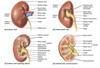
What are the different intrarenal structures?
- hilum
- sinus (typically filled w/ fat)
- pelvis
- calices
- cortex
- medulla
- pyramids
- columns
- papilla
- segmental a./v.
What is the location and constrictions of the ureters?
- location: run inferiorly from renal pelvis passing over pelvic brim and along lateral pelvic wall, running posterior to ducuts deferens (male) and uterine artery (female)
- constrictions (potential sites of obstructions): (1) junction of ureter w/ renal pelvis, (2) crossing pelvic brim, and (3) entering bladder wall
- pyelogram: x-ray of ureter enhanced w/ contrast media

What is the arterial supply to the kidneys and ureters?
renal arteries: segmental arteries
ureter:
- renal branches
- gonadal branches
- abdominal aorta branches
- iliac branches
- superior vesical branches
- pelvic branches: rectal, uterine, vaginal, inferior vesicular

What is the location, blood supply, and innervation of the suprarenal (adrenal) glands?
- right suprarenal gland: lies near right crus, right kidney, and IVC
- left suprarenal gland: lies near left crus, spleen, stomach, and pancreas
- blood supply: superior, middle, and inferior suprarenal arteries; corresponding veins
- innervation: preganglionic sympathetic from T10-L1; celiac plexus and abdominopelvic splanchnic nerves

What is the lymphatic drainage of the posterior abdominal wall?
- common iliac lymph nodes: from external/internal iliac nodes, drain into lumbar lymph nodes
- lumbar lymph nodes: run along both sides of IVC and aorta, receive from posterior abd/pelvic walls and organs, form lumbar lymphatic trunks
- pre-aortic lymph nodes: form intestinal lymphatic trunks draining from alimentary tract, liver, spleen, and pancreas
- all collected into cisterni chyli (beginning of thoracic duct)


