Gut Immunology (Shnyra) Flashcards
- the largest immune organ in the body
- consists of multi-follicular Peyer’s patches and isolated lymphoid tissue (ILT)
- cross-talk between host immune system and microbiota is critical for this organ and ILT development that in turn regulates the microbiota
gut-associated lymphoid tissue (GALT)

Describe the role of GALT in the host-microbiota mutualism in intestines:
- after birth, bacteria immediately colonizes neonatal intestine initiating events that affect development and maturation of mucosa and GALT
- GALT consists of isolated lymphoid follicles (ILFs) and Peyer’s patches (PPs)
- GALT is primary route by which body is exposed to Ags (microbial and diet)
Describe the role of ILFs in the host-microbiota mutualism in intestines:
- ILFs develop after birth in SI and LI, showing dynamic response of gut immune system to microbiota
- ILFs are single B-cell follicles that act as inductive site for IgA prod
- ILFs lack afferent lymphatic vessels and receive Ags directly from epithelial surface via Ag-transporting DCs
Describe the role of Peyer’s patches (PPs) in the host-microbiota mutualism in intestines:
- along w/ ILFs, make up GALT
- lack afferent lymphatic vessels and therefore receive Ags directly from epithelial surface via Ag-transporting DCs
- microbes also cross epithelium and enter PPs through M cells, from which they are endocytosed by DCs in subepithelial dome
- Ag-loaded DCs in the PPs interact w/ local lyphocytes to induce differententiation of T cells and T cell dependent B cell maturation in the germinal center to induce the development of IgA-producing plasma cells
- after the plasma cells hone into the lamina propria, they release secretable dimeric IgA for transport into the intestinal lumen
Describe the role of microbe-associated molecular patterns (MAMPs) in the host-microbiota mutualism in intestines:
- MAMPs recognized by pattern-recognition receptors (PRRs) on intestinal epithelial cells, DCs adjacent to crytopatches stim the recruitment of B cells and T cells, causing cryptopatches to develop into mature ILFs
- PRR-mediated recognition of MAMPs stim proliferation of intestinal epithelial cells in crypts, resulting in their increased depth and density of Paneth cells in SI
- MAMPs also stim intestinal epithelial cells for release of antimicrobial peptides (defensins)
How does the intestinal epithelium provide a barrier to exogenous materials?
- subpopulations of intestinal epithelial cells (IECs) are integrated into a continuous, single layer
- goblet cells: prod mucin, organized into a dense, more highly cross-linked inner proteoglycan gel and a less densely cross-linked outer mucous layer
- enterocytes (SI), colonocytes (LI), and Paneth cells (base of SI crypts): continually sense the microbiota (MAMPs) to induce prod of antimicrobial peptides (AMPs)
- secretory IgA (sIgA): maintains peaceful bacteria-host interaxn; does not active complement sys; does not active phagocytes in Fc-receptor dependent manner; resistant to proteolysis by peptidases prod in stomach, SI, and pancreas
- AMPs: contibute to mucosal host defense in GI
- defensins: prod by IECs, major class of AMPs in GI which represent innate immunity

How do bactericidal defensins protect against pathogens?
- high density/conc of defensins within inner mucous layer makes in largely impervious to bacterial colonization/penetration
- have clusters of positively charged AA side chains (pink) and hydrophobic AA side chains (green)
- polarity allows defensins to interact w/ microbial membranes that results in form of membrane “wormholes” or pores
- est that innate immune system provides protection against ~98% of pathogens that encountered by the body

Describe the role of adaptive immune responses within the GI:
- most commensal bacteria reside outside layer of mucus that covers IECs
- commensal and pathogenic bacteria that penetrate enterocyte epithelial layer are rapidly killed by Mφ in lamina propria
- bacteria can also penetrate specialized follicle-associated epithelium, containing M cells that lie over PPs, which are rapidly killed by Mφ; however some can be picked up by DCs
- DCs interact w/ T and B cells in PPs and/or migrate to mesenteric lymph nodes (LNs), and induce IgA producing plasma cells
- DCs loaded w/ commensal bacteria can traffic to mesenteric LNs, however the LNs function as a barrier, meaning loaded DCs cannot penetrate farther to reach systemic circulation
- following activation, Ag-activated B and T cellsleave mesenteric LNs through efferent lymph, enter the BS at the thoracic duct and hone back to intestinal mucosa

What is the role of Treg cells in the GI?
- recall: Treg cells are T cells w/ high affinity for self-Ags that express transcription factor Foxp3 and become natural T regulatory cells

What is the role of diet, environment, and genetics in gut microbiota?
- changes in these factors effect microflora
- balanced microbial composition results in symbiosis > immune regulation and homeostasis
- microbial imbalance results in dysbiosis > immune dysregulation and inflammation in susceptible host (genetics)
- dysbiosis can occur due to changes in diet and other environmental factors

- produced by colonic microbial fermentation of undigested or partially digested dietary fibers/carbs
- have broad effects on host immune system development
- examples: butyric acid (butyrate), propionic acid (propionate), acetic acid (acetate)
short-chain fatty acids (SCFAs)

What is the role of diet in the GI microbiota and immune system?
- diet shapes gut microbiota (structurally/functionally), microbiota adapts to promote nutrient processing
- microbiota and IS co-evolve: malnutrition affects both IS and microbiota
- undernutrition: a/w defects in innate/adaptive immunity
- recurrent enteric infections: predispose to nutrient deficiencies and impaired intestinal mucosal barrier function
- increase susceptibility to infection and worsening nutritional status
- microbiota acts as barrier to enteropathogen infection, the barrier may be disrupted by malnutrition

What is SCFAs role in cell differentation in the GI?
- SCFAs promote differentation of Treg and sIgA production
- acetate stim accumulation of IL-10 producing colonic Tregs
- butyrate either directly acts on Tregs or modulates DC function to enhance Treg-inducing ability
- capsular polysaccharide A (PSA) derived from B. fragilis and MAMPs can directly act on Tregs through TLR2 to promote Treg function by enhancing expression of effector molecules (IL‐10 and TGF‐β)
- SCFAs help support effective IgA-mediated response to gut pathogens and stim prod of mucus

What are the different types of food tolerances and what is the clinical/immunological relevance?
- immune tolerance: sustained immune unresponsiveness to self-Ags, beneficial Ags, and commensal bacteria
- oral tolerance: suppression of immune responses to Ags that have been administered by oral route
- failure to induce food tolerance to through to result in food allergy and celiac dz (most prevenlant food-induced pathology)
type of immune tolerance
immature lymphocytes specific for self Ags may encounter these self Ags in the generative lymphoid organs and are either:
- deleted (apoptosis)
- change BCR specificity (B cells only)
- develop into Treg cells
central tolerance

type of immune tolerance
mature self-reactive lymphocytes in peripheral tissue may be either:
- inactivated (anergy)
- deleted (apoptosis)
- suppressed by Treg cells
peripheral tolerance

What is the role of peripheral tolerance in the GI?
- central tolerance of T cells and nTreg cell differentiation require interaction of TCR w/ its cognate Ag in the thymus
- since the intestinal Ags are not available in the thymus, central tolerance cannot prevent responses against Ags in the lamina propria
- peripheral tolerance is needed to ensure tolerance to Ags such as food Ags and commensal organisms
What is the mechanism of oral tolerance?
- Mφ, DCs, and Treg cells play crucial role in OT
- DCs: take up Ags from intestinal lumen by sending cellular processes into lumen across epithelial barrier
- Mφ: transfer acquired Ags to DCs in lamina propria (LP)
- Ag loaded DCs move from LP to mLNs in a chemokine dependent manner
- in mLNs, DCs stim naive CD3+ T cells to differentiate into induced CD4+CD25+Foxp3+ Treg cells via release of retinoic acid (RA), TGF‐β, and indoleamine 2,3‐dioxygenase (IDO)
- RA: directly induced Treg-cell differentiation
- TGF-β: mediates Foxp3 upregulation in Treg differentiation
- IDO: exerts immunosuppresive functions causing anergy of effector T cells and induces proliferation of Treg cells

What are the different types of adverse food reactions?
toxic: food poisoning
non-toxic:
- non-immune mediated (food intolerance): pharmacological, enzymatic, psychosomatic, irritant
- immune mediated (food allergy): IgE mediated (type I), non-IgE mediated (type III/IV)
What are examples of non-immune mediated food reactions?
- absence of enzyme: needed to fully digest food (lactose intolerance)
- irritable bowel syndrome: chronic, can cause cramping, constipation, diarrhea
- food poisoning: toxins such as bacteria in spoiled food can cause severe GI sx
- recurring stress/psychological factors: reason not fully understood
What are examples of immune-mediated food rxns?
- food allergy and celiac dz: arise from specific immune reponse that occurs reproducibly on exposure to a given food
- sensitivity to food additives: sulfites used to preserve dried fruit, canned goods, and wine can trigger asthma attacks in sensitive people
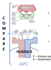
- celiac dz: chronic digestive condition triggered by gluten (protein in wheat/grains); has some features of a true food allergy, however ppl w/ celiac dz not at risk for anaphylaxis
What are the different types of immune-mediated food rxns?
- type I hypersensitivity (most common): development of IgE against food allergens; patients w/ this type of food allergy can be identified by measuring in vivo IgE-mediated skin rxn to food allergen or measuring specific IgE in serum/body fluids
- type III/IV hypersentivities: activation of macrophages by allergen-Ab complexes in FcγR‐dependent manner (type III) and activation of allergen-specific T cells (IV); delayed, can take up to 48 hr to develop, but still involve immune system
- genetic predisposition and environmental factors may abrogate oral tolerance and lead to this allergy
- affects ~5% of young children and 3-4% of adults in westernized countries
- increasing in prevalance
- can cause variety of sx/disorders involving skin, GI, and respiratory tracts
- can be attributed to IgE-mediated (type I) and non-IgE-mediated (type III/IV) mechanisms
food allergy
What is the IgE-mediated mechanism of food allergies?
- primary encounter and subsequent exposure
- allergic sensitization: when IgE-associated food allergies develop early in childhood
- allergen contact via GI tract, resp tract, and skin induces IgE production (primary sensitization) in genetically predisposed individuals
- repeated allergen contact activates allergen-specific T cell and induces IgE-dependent secondary immune response
- factors that affect integrity of epithelial barrier and extent of allergen digestion are important for primary sensitization and boosting of secondary responses
- after allergen ingestion/degradation, fragments and internalized from GI and distributed throughout body
- thus, allergic response develops not only in intestine, but also organs such as skin, resp tract, circulatory sys (which leads to anaphylaxis)