Motility of the GI Tract (Lopez) Flashcards
- one of the major activities taking place in the GI tract
- involves the contraction and relaxation of walls and sphincters of the GI tract
- rate of this process is important to control
motility

What are the functional layers of the GI tract?
- mucosal layer: muscularis mucosae consists of smooth muscle, its contractions change the shape and surface area of the epithelium
- submucosa
- muscle layers (muscularis propria): smooth muscle layers (circular and longitudinal), provide motility to the GI tract, contractions of this layer mix and circulate the content of the lumen and propel them through the GI tract
- serosa

What are the differences in function of the circular and longitudinal muscles of the muscularis propria layer?
- circular muscle: contraction decreases diameter of the segment
- longitudinal muscle: muscle contractions decreases length of the segment
- electrophysiological event within the GI that entails depolarization and repolarization of the membrane potential (not an AP)
- in part, responsible for the coordinated contractions and relaxations of the musculature necessary for the efficient grinding, mixing and transportation of the food we ingest
- generated and propagated by a class of pacemaker cells called the interstitial cells of Cajal, which also act as intermediates between nerves and smooth muscle cells
slow waves

- periodic contractions followed by relaxation
- e.g.: esophagus, stomach (antrum), SI, and all tissue involved w/ mixing and propulsion
phasic contractions

- contractions maintained at a constant level w/o regular periods of relaxation
- e.g.: stomach (orad), lower esophageal, ileocecal, and internal anal sphincters
tonic contractions

What is the relationship between slow waves, action potentials, and contractions within smooth muscle?
- the greater the # of APs on top of the slow wave, the larger the contraction
- neural and hormonal activity modulate generation of APs and strength of contraction
- the mechanical response follows the electrical response

What is the relationship between the NT’s, Ach and NE, and slow waves and action potentials?
- ACh: increases the amplitude of slow waves and the # of Aps
- NE: decreases the amplitude of slow waves

What is the role of the enteric nervous system (submucosal plexus and myenteric plexus) in GI motility?
- submucosal plexus (Meissner’s): located in submucosa, mainly controls GI secretions and local blood flow
- myenteric plexus (Auerbach’s): located between longitudinal and circular layers, mainly controls GI movements

- the pacemaker cells of the GI smooth muscle
- generate and propagate slow waves
- slow waves occur spontaneously within these cells and spread rapidly to smooth muscle cells via gap junctions
- electrical activity in these cells drives the frequency of contractions
- these cells are located in the pacemaker region of the GI (myenteric plexus) and smooth muscle (intramuscular)
interstitial cells of Cajal (ICC)

Describe mastication in terms of innervation:
- most of the muscles of mastication are innervated by the motor branch of CN V
- act of mastication is both voluntary and involuntary
- controlled by nuclei in the brain stem
- caused by a chewing reflex

What are the phases of swallowing and are they voluntary/involuntary?
swallowing initiated voluntarily in the mouth, and after is under involuntary reflex control thereafter
- oral phase (voluntary): initiates swallowing process
- pharyngeal phase (involuntary): soft palate is pulled upward > epiglottis moves > UES relaxes > peristaltic wave of contractions initiated in pharynx > food is propelled through open UES
- esophageal phase (involuntary): control by swallowing reflex (primary peristalsis) and ENS; secondary peristalsis can occur through esophageal distention

What part of the brain controls the swallowing reflex and how is it initiated?
- involuntary swallowing reflex is controlled by the medulla
- food in pharynx > afferent sensory input via vagus/glossopharyngeal N. > swallowing center (medulla) > brainstem nuclei > efferent input to pharynx

What are the differences between primary and secondary perstaltic waves?
- primary peristalsis: continuation of pharyngeal peristalsis (swallowing reflex), controlled by the medulla, cannot occur after vagotomy
- secondary peristalsis: occurs if primary wave fails to empty esophagus or if gastric contents reflux into esophagus (esophageal distention), medulla and ENS are involved, can ocur in absence of oral and pharyngeal phases, occurs even after vagotomy

What occurs with the pressure along the esophagus as food passes through it?
- changes in pressure occur
- generally, pressure increases where the food bolus is located
- once the bolus passes below the level of the diaphragm, pressure decreases along the esophagus where the bolus is located

What challenges are posed from the intrathoracic location of the esophagus and how are these problems solved?
two problems:
1) keeping air out of the esophagus at the upper end
2) keeping acidic gastric contents out of the lower end
problems solved:
- UES and LES are closed, except when food bolus is passing from pharynx to esophagus or from esophagus to stomach
- condition that involves impaired peristalsis, incomplete LES relaxation during swallowing (LES stays mostly closed during swallowing, resulting in backflow of food), and elevation of LES resting pressure
- etiology: decreased # of ganglion cells in myenteric plexus, degeneration preferentially involves inhibitory neurons producing NO/VIP, or damage to nerves in esophagus preventing it from squeezing food into stomach
- presentation: backflow of food in the throat (regurgitation), difficulty in swallowing both liquid and solids, heartburn, chest pain
achalasia
- condition that involves changes in the barrier between the esophagus and stomach (e.g. LES relaxes abnormally or weakens)
- etiology: motor abnormalities that result in abnormally low pressure in the LES, or increased intragastric pressure (e.g. after a large meal, during heavy lifting, during pregnancy)
- persistent reflux and resulting inflammation leads to this condition
- presentation: backwash of acid, pepsin and bile in the esophagus can lead to heartburn and acid regurgitation; also, GI bleeding, esophagitis, scar tissue in esophagus (Stricture of esophagus), and Barrett’s esophagus
GERD
What are the regions of the stomach based upon differences in motility?
- anatomical divisions: fundus, body, and antrum
- functional regions: orad and caudad
- orad: receptive relaxation
- caudad: mix and digestion

What are the muscular layers and innervations of the stomach?
- 3 layers of muscle: circular, longitudinal, and oblique
- extrinsic innervations: parasympathetic and sympathetic
- intrinsic innervations: myenteric and submucosal plexuses (ENS)
What is the function of the orad region of the stomach?
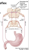
- receptive relaxation: decreased pressure and increased volume of the region; vagovagal reflex (controls contraction of the GI muscle layers in response to distension of the tract by food; also allows for the accommodation of large amounts of food in the GI tract)
- orad region exhibits minimal contractile activity (little mixing of ingested food occurs there)
- CCK decreases contractions and increases gastric distensibility
(CCK: hormone which is secreted by cells in the duodenum and stimulates the release of bile into the intestine and the secretion of enzymes by the pancreas)

What is the function of the caudad region of the stomach?
- mix and digestion
- primary contractile event is peristaltic contraction (mid stomach > pylorus)
- contractions increase in force and velocity as they approach the pylorus
- max frequency is ~3-5 waves/min
- retropulsion also occurs here (gastric contents propelled back into stomach for further mixing)

- process where most of the gastric contents are propelled back into the stomach for further mixing and reduction of particle size
- peristaltic waves move from mid stomach to antrum, which in turn closes the pylorus
- closing of pylorus causes this to occur
retropulsion

What is the sequence of gastric motility movements?
- stomach fills, peristaltic waves from antrum move toward pylorus, most of gastric contents are pushed back into body of stomach
- first wave fades out, stronger wave originates at incisure and squeezes gastric contents in both directions
- pylorus opens as secon wave approaches, duodenal bulb is filled and some contents pass into second portion of duodenum, third wave starts just above incisure
- pylorus closes again, third wave fails to evacuate contents, fourth wave starts higher on body of stomach, duodenal bulb may contract or remain filled as peristaltic wave originating just beyond it empties second portion of duodenum
- peristaltic waves are originating higher on body of stomach and gastric contents are evacuated intermittently, contents of duodenal bulb are pushed passively into second portion as more gastric contents emerge
- 3-4 hours later stomach is almost empty, small peristaltic wave empties duodenal bulb w/ some reflex into stomach, reverse and antegrade peristalsis occur in duodenum


