Development of GI System (Brauer) Flashcards
(43 cards)
- cranio-caudal folding of the embryo creates the primitive gut tube which is comprised of these structures:
- around week 4, the midgut’s connection to the yolk sac narrows, creating this structure:
- cranio-caudal folding of the embryo creates the primitive gut tube which is comprised of these structures: foregut, midgut, hindgut
- around week 4, the midgut’s connection to the yolk sac narrows, creating this structure: vitelline duct

GI structures derived from endoderm:
- mucosal epithelium
- GI glands (except lower 1/3 of anus)

GI structures derived from splanchnic mesoderm:
- connective tissue
- vasculature
- smooth muscle wall

GI derivatives derived from ectoderm:
- enteric ganglia, nerves, and glia (via neural crest cells)
- epithelium of lower 1/3 anus

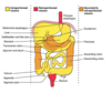
What are the structures of the foregut and what is the arterial supply?
- structures: esophagus, stomach, liver, gallbladder, pancreas, upper duodenum
- BS: celiac trunk

What are the structures of the midgut and what is the arterial supply?
- structures: lower duodenum, jejunum, ileum, cecum, appendix, ascending colon, proximal 2/3 of transverse colon
- BS: superior mesenteric artery (SMA)

What are the structures of the hindgut and what is the arterial supply?
- structures: distal 1/3 of the transverse colon, descending colon, sigmoid colon, rectum, upper anal canal
- BS: inferior mesenteric artery (IMA)

What is the function of peritoneum within the abdomen and what are the different types of peritoneum?
- peritoneum: thin membrane that lines the abdominal and pelvic cavities, and covers most abdominal viscera; composed of layer of mesothelium supported by a thin layer of connective tissue
- parietal peritoneum: the portion that lines the abdominal and pelvic cavities (also known as the peritoneal cavity)
- visceral peritoneum: covers the external surfaces of most abdominal organs, including the intestinal tract

GI structures derived from dorsal mesentary:
- greater omentum: gastrosplenic, gastrocolic, splenorenal ligaments
- small intestine mesentery
- mesoappendix
- transverse mesocolon
- sigmoid mesocolon

GI structures derived from ventral mesentery:
- lesser omentum: hepatoduodenal and hepatogastric ligaments
- falciform ligament of liver
- coronary ligament of liver
- triangular ligament of liver
- organs that are suspended by mesentery
- stomach, tail of pancreas, first five cm and the fourth part of the the duodenum, jejunum, ileum, cecum, appendix, transverse colon, sigmoid colon, and upper third of the rectum
intraperitoneal organs

- organs that are excluded from peritoneal cavity
- mnemonic: SAD PUCKER
Suprarenal (adrenal) gland
Aorta/IVC
Duodenum (2nd and 3rd part)
Pancreas (except tail)
Ureters
Colon (ascending and descending)
Kidneys
(o)Esophagus
Rectum
retroperitoneal organs

- organs that were initially suspended within mesentery that later fused w/ body wall
- ascending/descending colon, duodenum, bulk of pancreas
secondarily retroperitoneal organs
Mnemoic SAD PUCKER is used to recall:
retroperitoneal organs
S - suprarenal (adrenal) glands
A - aorta/IVC
D - duodenum (2nd and 3rd parts)
P - pancreas (except tail)
U - ureters
C - colon (ascending/descending)
K - kidneys
E - (o)esophagus
R - rectum

GI structures derived from foregut:
- esophagus
- stomach
- liver and gallbladder
- pancreas
- upper duodenum (proximal bile duct)
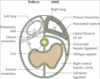
When the stomach (foregut) rotates during development, what anatomical changes occur and what structures develop?
- stomach is attached to body wall by peritoneum (mesentery) both ventrally and dorsally
- stomach rotates 90 degrees wherein left side moves ventrally and right side moves dorsally
- ventral mesentery > lesser omentum (located on lesser curve of stomach)
- dorsal mesogastrium > greater omentum (located on greater curve of stomach)
- lesser sac located behind stomach
- vagus nerve (LARP): left vagus nerve > anterior; right vagus nerve > posterior

How do the greater and lesser omentums develop?
- ventral mesentery > lesser omentum
- dorsal mesogastrium > greater omentum
- lesser omentum located between lesser curve of stomach and liver
(epiploic foramen: opening to lesser sac between liver and top of duodenum)
- greater omentum grows downward in a double fold in front of transverse colon
- greater omentum superior to transverse colon comes together w/ transverse mesocolon (attaches colon to pancreas and body wall) and duplicated layers are absorbed > greater omentum becomes stuck to transverse colon while also hanging below it

How do the greater and lesser sacs develop?
(side note: liver develops in ventral mesogastrium and spleen develops in dorsal mesogastrium)
- rapid and large growth of liver causes lesser sac to develop
- lesser sac: located behind lesser omentum, stomach, and gastrocolic ligament (part of the greater omentum)
- greater sac: larger portion of the peritoneal cavity; divided by the transverse mesocolon into supracolic and infracolic compartments
- epiploic foramen: opening within lesser omentum from greater sac to lesser sac

- thickened muscle causing narrowing of opening between stomach and duodenum
- etiology: faulty migration of neural crest cells > ganglion cells of enteric nervous system not properly populated which causes inability of sphincter to relax; or narrowing of pyloric lumen due to hypertrophy of muscularis externa in this region
- presentation: occurs within a few months after birth, palpable mass (“olive”) at right costal margin, projectile non-bilious vomiting after feeding, fewer and smaller stools, failure to gain weight (may lose weight)
- incidence: 1:500
hypertrophic pyloric stenosis

How does the liver develop?
- during week 4, begins as hepatic diverticulum from gut endoderm
- connection of diverticulum to foregut > common bile duct
- endoderm > hepatocytes, bile ducts, and hepatic ducts
- splanchnic mesoderm > stromal, Kupffer, and stellate cells
- takes over hematopoiesis in utero by week 10
- congenital anomalies are rare

Describe the development of the gallbladder and bile duct:
- around week 4, begins as cystic diverticulum (outgrowths from cystic endoderm)
- gallbladder develops as a secondary out-pouching from hepatic diverticulum (common bile duct) that grows into ventral mesentery, the connection between the two is the cystic duct
- around the end of week 5, recanalization of bile duct occurs
- around the end of week 6, the common duct and ventral pancreatic bud rotate 180° clockwise around the duodenum so that the gallbladder remains situated inferior to the liver
- beginning week 12, bile is formed by hepatic cells

- blockage of the ducts that carry bile from the liver to the gallbladder
- both congenital and adult forms of condition
- etiology: obliteration of bile duct, or inflammation replaces duct w/ fibrotic tissue
- presentation: immediate onset of progressive jaundice in infants, white/clay colored stool, dark urine, poor prognosis (12-19 month lifespan)
- incidence: extrahepatic (1:15,000) and/or intrahepatic (1:100,000)
biliary atresia

Describe the development of the pancreas:
- around week 4, begins as two additional endodermal buds sprouting from the foregut, inferior to cystic diverticulum
- ventral pancreatic bud derivatives: uncinate process (grows into ventral mesentery)
- dorsal pancreatic bud derivatives: pancreatic head, body, and tail (grows into dorsal mesentery)
- buds develop w/ both exocrine and endocrine portions
- around week 5, ventral pancreas migrates around posteriorly to fuse w/ dorsal pancreas (ventral and dorsal ducts also fuse)
- ventral pancreatic duct > main pancreatic duct (connection to duodenum)
- dorsal pancreatic duct > accessory pancreatic duct

- developmental variation within pancreas where there is an additional pancreatic duct (Duct of Santorini) that occurs within 33% of people
- can be functional or non-functional and may open separately into the second part of the duodenum which is dorsal and usually (in 70%) drains into the duodenum via the minor duodenal papilla
- in the other 30% it drains into the main pancreatic duct, which drains into the duodenum via the major duodenal papilla
- the main pancreatic duct and this additional duct both eventually—either directly or indirectly—connect to the second part (‘D2’, the vertical segment) of the duodenum
accessory pancreatic duct
(left side of photo)


