Female Reproductive Histology (Dennis) Flashcards
What are the 2 coexisting cycles that occur within the menstrual cycle?
- ovarian cycle: several ovarian follicles undergo folliculogenesis in preparation for ovulation
- uterine cycle: concurrent cycle, where the endometrium prepares for implantation
What structures are a/w the ovaries?
- ovaries are lined by ovarian surface epithelium (OSE): simple cuboidal epithelium, embryonic source of granulose cells and stromal cells > comprise the growing follicles; overlying layer of dense CT capsule (tunica albuginea)
- contain a peripheral cortex w/ deep medulla: cortex is CT and ovarian follicles; medulla is CT, interstitial cells, neurovasculature, and lymphatics via the hilum

- located in the cortical stroma
- contain a single oocyte
- follicular/granulosa cells surround occyte and support its growth
- early stages of oogenesis occur during fetal life
- oocytes present at birth remain arrested in meiosis I
ovarian follicles

What are the phases of folliculogenesis?
(select follicles undergo cyclic growth and maturation, phases do not occur simultaneously)
1) follicular phase
2) ovulatory phase
3) luteal phase
What are the different types/stages of primary ovarian follicles?
- primordial follicles: numerous throughout the cortex, ~25 μm in diameter, surrounded by simple squamous layer of follicular/pregnulosa cells
- primary follicles: simple squamous granulosa cells > simple cuboidal layer of granulosa cells; basal lamina separates the granulosa cells from the stroma of the ovary; zona pellucida begins to assemble, separates primary oocyte from granulosa cells
- late primary follicles: stratified granulosa cells communicate through gap junctions; follicle is still avascular and has basement membrane

- the type of follicle that forms when granulosa cells begin to secrete follicular fluid
- fluid accumulates in small spaces, Call-Exner bodies, which eventually enlarge and combine
- granulosa cells reorganize themselves around a larger cavity, the antrum
- stromal cells form separate thecal layers
secondary follicle

What are the different types of thecal layers and how are they formed?
(stromal cells proliferate into a stratified cuboidal epithelium, the theca)
- theca interna: vascularized cell layer adjacent to the basal lamina supporting granulosa, produces androstenedione > estradiol
- theca externa: fibrous cellular layer c/w ovarian stroma

How do mature (Graffian) follicles develop?
- antrum accumulates more fluid, reaches its maximum size (2 cm)
- thecal layers thicken
- buldges at the surface of the ovary are visible w/ ultrasound

What are the different types of granulosa cells within a mature follicle?
(granulosa cells thin out and are segregated by fluid)
- mural granulosa cells: line follicular wall, actively synthesize and secrete estrogen, prod follicular fluid
- cumulus oophorus: anchor primary oocyte to follicle, nutrient delivery channel
- corona radiata: granulosa cells anchored to ZP

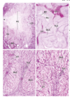
What type of follicle is this? Identify structures in the image:

primordial follicles
- SE: surface epithelium
- TA: tunica albuginea
- O: oocyte
- arrows: epithelial follicular cells

Identify structures in the following image:

- PF: primordial follicles
- UF: primary follicles
- G: follicular cells
- O: oocyte

What type of follicle is this?

secondary follicle
Identify structures in the following image:

- A: antrum
- AF: atretic follicle
- F: follicle cells, primordial
- FC: follicle cells
- GC: granulosa cells
- GEp: germinal epithelium
- GF: growing follicles
- N: nucleus of oocyte
- PF: primordial follicles
- TA: tunica albuginea
- TI: theca interna
- X: oocyte w/ only cytoplasm
- ZP: zona pellucida
- arrowhead: follicle cells seen en face

Describe the process of follicular atresia:
- during menstrual cycle, one follicle becomes dominant follicle
- the other primary and antral follicles undergo atresia (failure of follicle to ovulate)
- apoptosis is the mechanism: ensures regression of follicle w/o causing inflammatory response; glassy membrane (thick folded basement membrane material)

What occurs during the ovulatory phase of ovarian cycle?
LH surge causes:
- primary oocyte completes meiosis I > secondary oocyte (arrested at metaphase II)
- oocyte undergoes ovulation and enters oviduct
- mural granulosa cells and theca interna repair OSE damage following follicle rupture

What occurs during luteinization of the luteal phase of ovarian cycle?
- thecal cells differentiate to form corpus luteum, promotes endometrial changes that support implantation: mural granulosa cells > granulosa lutein cells; theca interna cells > theca lutein cells
- granulosa lutein cells: hypertrophic, steroid-secreting; secrete progesterone and estrogen w/ FSH and LH stimulation
- theca lutein cells: produce androstenedione and progesterone w/ LH stimulation

Identify structures in the following image:

- BV: blood vessel
- CT: connective tissue
- FC: former follicular cavity
- GLC: granulosa lutein cells
- TC: granulosa cells transforming into corpus luteum cells
- TLC: theca lutein cells

What occurs during luteolysis of the luteal phase of the ovarian cycle?
if fertilization occurs:
- CL continues to prod progesterone and estrogen under stimulatory action of human chorionic gonadotropin (hCG) from trophoblast layer
if fertilization does not occur:
- CL begins involution stage ~14 days after ovulation
- luteolysis (regression of CL) leads to formation of corpus albicans (scar of CT, type I collagen w/ few fibroblasts) which forms at the site of CL after involution, gradually becomes very small

What is the histological structure of the uterine tube?
- walled set of ducts that provide fertilization microenvironment and transport embryo to uterus
- mucosal layer: simple columnar epithelium w/ lamina propria of loose CT; ciliated cells and secretory peg cells are sensitive to estrogen signaling and will increase in size
(mucosal folds are larger in areas of the ampulla compared to areas of the isthmus)
- smooth muscle layer: inner circular-spiral layer and outer longitudinal layer, peristaltic contractionandciliary activity propel oocyte/zygote toward uterus
- serosa layer w/ large blood vessels


- concurrent cycle w/ the ovarian cycle
- endometrium prepares for implantation
- if fertilization does not occur, endometrium is shed and menstruation occurs as a new menstrual cycle begins
uterine cycle
What is the histological structure of the uterus?
- perimetrium: serosa covering posterior surface and part of anterior surface (remainder is adventitia)
- myometrium: contains poorly defined smooth muscle; central, circular layer that is thick w/ blood vessels is the stratum vasculare; outer and inner layers contain longitudinally or obliquely arranged fibers
- endometrium: epithelium is simple columnar w/ simple tubular glands; functional layer is lost during menstruation, supplied by spiral arteries; basal layer is retained during menstruation


