Part 14 Flashcards
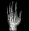
Patient is a 60 year old male has and has been complaining of pain, unexplained weight loss and recurrent respiratory infections. You take this xray and labs. Elevated ESR and M spike. What are the findings? Diagnosis?

Osteoporosis/osteopenia, soap bubble appearance
Multiple myeloma
Diagnosis?

Multiple myeloma
Findings? Diagnosis?

Sunburst periosteal reaction, dense sclerotic lesion at the proximal aspect of the humerus in the metaphyseal region, Codman triangle
Osteosarcoma
Findings? Diagnosis?

Cumulus cloud like density
Osteosarcoma
Findings? Diagnosis?

Stippled calcifications, popcorn like density, round oval area of demineralization, lytic destruction of proximal humerus
Chondrosarcoma
Findings? Diagnosis?

Onion skin appearance in the diaphysis of the femur
Ewing’s sarcoma
Findings? Diagnosis?

Geographic, eccentric, metaphyseal/epiphyseal, subarticular, soap bubble appearance, lytic, cortical thickening
Giant cell tumor
Findings? Diagnosis?

Sessile, metaphyseal bony exostosis
Solitary osteochondroma
Findings? Diagnosis?

Pedunculated, cortical rim, coat hanger exostosis, cauliflower cap
Solitary osteochondroma
Findings? Diagnosis?

Multiple Osseous stalks of the tibia, fibula and femur
Hereditary multiple exostosis
Findings? Diagnosis?

Impaired bone growth, short ulna, bowing of radius, subluxation of the radioulnar joint
Madelung deformity
Findings? Diagnosis?

Geographic, central, oval radiolucency with long axis parallel to bone
POSSIBLE FALLEN FRAGMENT SIGN, OPEN GROWTH PLATES
Solitary bone cyst
Findings? Diagnosis?

Lytic , eccentric lesion in the metaphysis, soap bubble appearance
GROWTH PLATES, NOT SUBARTICULAR
aneurysmal bone cyst
Findings? Diagnosis?

Radiolucent, expansile lesion
Osteoblastoma
Findings? Diagnosis?

Large oval lesion with a sclerotic border, >1cm
Osteoblastoma
This patient has pain in the proximal tibia and he can take ibuprofen and it gets better. What are the findings and diagnosis?

Oval shaped Nidus <1cm in size with a dense sclerotic border in the metaphysics of the bone
Osteoid osteoma
Findings? Diagnosis?

Well circumscribed round extremely dense lesion around 2cm in size in the frontal bone
Osteoma
Findings? Diagnosis?
Round/oval lucency with fine marginal lines, centrally located, endosteal scallop ingredients, stippled or punctuate calcifications
Enchondroma
Findings? Diagnosis?

Multiple enchondromas
Ollier’s disease
Diagnosis?

Maffucci syndrome
Findings? Diagnosis?
Exaggerated vertical trabeculations (honeycomb appearance), posterior bulge of cortex
Hemangioma
Findings? Diagnosis?

Round eccentric lesion in apophysis or epiphysis, well defined sclerotic margin
Chondroblastoma
Findings? Diagnosis?

Round lyrics lesion 1-2cm in diameter, well defined sclerosis margin, eccentric and metaphyseal
Fibrous cortical defect/fibrous xanthoma
Findings? Diagnosis?

Round lyrics lesion >2cm in diameter, well defined sclerosis margin, eccentric and metaphyseal
Non ossifying fibroma/fibrous xanthoma
Findings? Diagnosis?

Well defined geographic lysis, cotton wool appearance
Paget disease
Findings? Diagnosis?

Thickening of iliopectineal line and cortical lines of public bones (rim sign, brim sign), cortical Thickening, Thickening and disorganization of trabeculae, acetagular protrusion
Paget disease
Findings? Diagnosis?

Ground glass appearance in proximal femur
Fibrous dysplasia
Sign? Diagnosis?

Shepard’s crook deformity
Paget disease
Sign? Diagnosis?

Blade of grass/candle flame deformity
Paget disease
Sign? Diagnosis?

Banana fracture
Paget disease
Sign? Diagnosis?

Sabre shin deformity
Paget disease
Findings? Diagnosis?

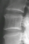
enlarged vertebra, enlarged cortices, picture frame appearance, accentuated trabeculation, thickened endplates
Paget’s disease
