Ortho Radiology 2 Flashcards
Midforearm Fractures
- Which parts of the forarm are unstable?
Isolated ulnar fractures
- If displaced less than 50% bone diameter and less than 10 degrees angulation what should we do?
3 YO that stops weight bearing = R/O what?
1.
- Radius
- ulna,
- midshaft are unstable
2. Short arm cast 2 weeks then functional splint 2-6 weeks
septic arthritis
What will an xray of a Frykman I AP view show?
3

- What percent of all distal radial fractures are treated?
- What are the normal distal radius findings on XR? 3
- 1/6 all fractures treated
- XRs
- Radial inclination
- Radial height- 1 cm
- Volar tilt- normal 10 degrees
- What is this?
- What signs will you find to support this Dx? 2

- scaphoid fracture- proximal part becomes avascular
2.
- anatomical snuff box tenderness and
- proximal part dies- scaphoid fracture
Scaphoid fractures
- XR of wrist: which kinds?
- Look for widening of what? at what value?
- High risk for what? Greatest risk of this where?
- What may be a reason that you initially miss this?
- XRs of wrist
- PA,
- lateral
- scaphoid view (AP with 30 degress of supination and ulnar deviation) - Look for widening of scapholunate distance (>3 mm)
- High risk of nonunion: Greatest at proximal pole
- XRs may be normal initially
- What is scapholunate dissociation?
- Signs? 3
- scaphoid ligament lunate is torn
2.
- tenderness to palapation on dorsal radial side of wrist
- could have click with movement
- Terry Thomas sign

Scaphoid Fractures
- Repeat the Xray when?
- Other imaging? 3
- Repeat XR in 10-14 days
2.
- Bone Scan
- MRI
- CT
- Benefits of a bone scan with scaphoid fractures? 2
- What would a CT help with? 2
1.
- More cost effective than MRI
- Can show uptake in 72 hours
2. Help see fracture line and displacement
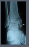
What is this?

Buckle fracture- pinching out on the outside like a water bottle
- What does this show?
- What is this fracture characterized by? 2
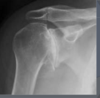
- Each of these anteroposterior radiographs shows an intrarticular fractures of the 1st metacarpal.
- the Type I or “Bennett’s” fracture of the proximal 1st metacarpal illustrated here is characterized by
- its articular involvement and
- the persistent attachment of the a volar fragment to the trapezium
- Where is the fragment that is dislocated in a Bennet’s fx?
- by what?
- Management?
Intra-articular fx through base of first metacarpal bone
- Large distal fragment dislocated radially and dorsally by
- the abductor pollicus longus muscle
- ORIF
Metacarpal Fractures
Thumb
- Bennett’s is a fracture combined with what? 2
- Whats a Rolando fracture?
- If extra-articular fracture how can we manage?
- What are acceptable limits for thumb for angulation and rotation?
- Bennett’s- fracture combined with
- subluxation or
- dislocation of metacarpal joint - Rolando fracture- T or Y shaped fracture involving joint surface (comminuted)
- If extra-articular fracture- can consider closed reduction and thumb spica cast for 4-6 weeks
- Less than 25 degrees angulation and rotation are acceptable limits for thumb
Would you fix this?
How should you check this?

You have to check rotation
If rotation if affected you need to fix if not you dont
make a fist and pinky dives underneath or sticks out
- What is this?
- How can you tell?
- Describe how this injury can progress with further trauma?
- If only one is broken how can you treat?

- Boxer’s fracture
- Nondisplaced and minimally angulated, neck
- boxers fracture- break 5th metacarpel and the next time they punch it they break the 4th. if they keep going they will keep breaking metacarpals
- with one you can probably just treat in a cast
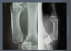
What is this?

Shaft, proximal transverse fracture
What does this show?

This anteroposterior radiograph shows a displaced fracture of the base of the 2nd metacarpal.
Metacarpal Fractures
- What should we examine for?
- How should we instruct the pt to do this?
- XRs?
- Examine for rotational malalignment
- Pt to make a fist
- XRs
- 3 view
Transverse fractures of the proximal phalanx are best seen on what kind of radiographs?
Lateral

How would you describe this fracture?
3

- transverse fracture of the proximal phalanx (white arrow).
- slight angulation of the two major fragments and
- the diastasis on the side adjacent to the arrow.
What kind of fracture is this?

This anteroposterior radiograph shows an oblique fracture of the proximal phalanx (white arrow). Oblique fractures may be stable if the periosteal sleeve is intact
What will heal faster: a transverse or oblique fx?
oblique: more surface area
What does this show?

The radiographs reveal a nondisplaced, nonangulated fracture (white arrow) of the proximal phalanx that does not appear to involve the proximal interphalangeal joint.
What kind of fx is this?

This AP x-ray shows a condylar fracture at the head of the proximal phalanx. These fractures most often involve one condyle and are unstable.
What is this?

The radiographs above show a fracture of the cancellous bone at the tip of the distal phalanx, often referred to as a tuft fracture. These fractures are generally stable and managed conservatively.
Have a subunguil hematoma- poke it? maybe