Opthalmology Anatomy Flashcards

1
Q
A

2
Q
A

Orbital rim

3
Q

Thin part of orbital rim?

What are they at risk of?

Complicatons of this?

A

Medial wall + orbital floor

* affected by orbital blowout fracture

* Comps: paraesthesia of skin of the face

4
Q

How do eyelids protect the eyes? (4)

What do they contain? (4)

A

* Protect eyes: outer skin, inner conjunctiva, eyelashes, glands

* Contain: tarsal plate (maintains shape), meibomian glands (secrete lipids), obicularis oculi (CN7), levator palpabrie superioris (CN3, elevates upper eyelid)

5
Q
A

6
Q

Lacrimal gland innervation?

Function?

A

CN7 (parasympathetic)

* produces tears

7
Q

Lacrimal apparatus?

A

* Lacrimal gland produces tears

* Pushed towards medial triangle

* drains through lacrimal puncta

* eventually reaches inferior meatus of nasal cavity

8
Q
A

9
Q

Layers of the eye? (3)

A

Fibrous - outer layer

* Sclera - white, fibrous

* Cornea - refraction

Uvea (vascular layer) - middle

* Iris

* Ciliary body - controls iris, shape of lens + aqueous humor secretion

* choroid - nutrition and gas exchange

Retina (photosensitive) - inner layer

10
Q

Where is anterior segment of the eye?

Divided into? (2)

A

In front of lens

* anterior chamber = between cornea and iris, contains squeous humor

* Posterior chamber = between iris and suspensory ligaments, contaisn aqueous humor

11
Q

Function of ciliary body of the eye? (3)

Where is it found?

A

Function

* controls iris

* shape of lens

* aqueous humor secretion

Found in uvea (middle layer)

12
Q

Posterior segment of the eye?

Function?

A

Posterior segment is behind the lens, and makes up 2/3rds of the eye

* contrains vitreous body which secretes virteous humor (common place for “floaters”)

13
Q

Describe circulation of aqueous fluid (4)

A

1) ciliary body

* secrete aqueous humor

2) aqueous circulates within posterior chamber and nourishes lens
3) squeous passes through pupil into anterior chamber, nourishes cornea
4) aqueous reabsorbed into scleral venous sinus (canal of Schlemm) at irodocorneal angle

14
Q

Arterial supply to eye?

A

Opthalmic artery from internal carotid artery

15
Q

What does the opthalmic artery divide into? (2)

Complication?

A

* Ciliary arteries + central artery of the retina

It is an end artery - cannot maintain tissue if occlusion occurs

16
Q

Venous drainage of the eye? (2)

A

* Superior opthalmic vein drains into cavernous sinusvia superior orbital fissure

* Inferior opthalmic vein drains into superior opthalmic vein

17
Q

Danger triangle of the face?

A

Infection in this area of the face can drain back into cavernous sinus and cause cavernous sinus thrombosis

18
Q

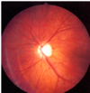
Parts of the retina? (4)

A

Fundus

*posterior area where light is focused

Optic disc

* point of CN 2 formation

* only point of entry/exit of blood vessels + axons of CN2

* blind spot!!

Macula

* greatest density of cones

Fovea

* centre of macula

* area of most cute vision

19
Q

Layers of the retina (from posterior to anterior)? (3)

Where do they retinal veins and arteries lie?

A

1) photoreceptors
2) ganglion cells
3) axons of ganglion cells

Retinal veins and arteries lie aterior to retina

20
Q

Where is the blind spot of the eye?

Why?

A

Optic disc

* there are no photoreceptors in the optic disc

21
Q
A

22
Q

What does interruption of flow in retinal artery branch/vein cause?

What about interruption of flow in central artery (end artery)?

A

* loss of an area of visual field corresponding to area of ischaemia

* monocular blindness

23
Q

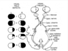
Where is light from objects in the right visual field processed?

Lower visual field?

A

* Left primary visual cortext

* upper part of visual cortext

24
Q

Extrinsic muscles of the eye? (6)

A
25
What is different about the superior oblique muscle of the eye?
It passes through a pulley (the trochlear)
26
Functions of the external muscles of the eye? (6)
\* Lateral rectus = laterally \* Medial rectus = medially \* Superior rectus = up and in \* Inferior rectus - down and in \* Superior oblique = down and out \* inferior oblique = up and out
27
Innervation of extrinsic muscles of the eye? (3)
\* Lateral rectus = abducent nerve \* Superior oblique = troclear nerve \* All others = CN III (occulomotor)
28
Sensory innervation of the eye?
Trigeminal nerve CN V1 (opthalmic) = upper eyelid, cornea, conjunctiva, tip + bridge of nose CN V2 (maxillary) = lower eyelid, maxilla, ala of the nose, upper lip CN V3 (mandibular) = mandible (except ridge of mandible, which is supplied by C2, 3)
29
Explain the blink (corneal) reflex? (4)
\* AP's conducted from cornea via CN V1 branches to trigeminal ganglion \* Then along CN V to pons \* CNS connections between CN V and CN VII \* AP's conducted via CN VII to obicularis oculi
30
Route of sympathetic axons? (4)
\* Originate from autonomic centres in brain \* Exit spinal cord with T1 - L2 spinal nerves (thoracolumbar outflow) \* Pass into sypathetic chains + **all** spinal nerves \* Pass into splanchnic nerves to supply organs
31
Function of splanchnic nerves? Types? (2)
Relay sympathetic nerves to organs \* Cardiopulmonary splanchnic nerves - heart + lungs \* Abdominopelvic= abdominal and pelvic organs
32
How are sympathetics relayed to the eyes?
3 cervical ganglia = superior, middle and inferior
33
How are sympathetic nerves carried to the eyes?
\* Travel from sympathetic trunk and synapse in superior cervical sympathetic ganglion \* Post-synaptic axons enter internal and external carotid nerves \* pass onto surface of internal and external carotid arteries \* carried to organs of the head \* opthalmic artery carries sympathetic axons to orbit (no splanchnic nerves involved)
34
Parasympathetic outflow to eye?
Craniosacral outflow \* CN3 + 7
35
Route of CN III (occulomotor)? What does it supply? (3)
Connects with CNS between midline and pons \* passes thru cavernous sinus \* exits via superior orbital fissure Supplies \* somatic motor to superior, medial, inferior rectus + inferior oblique \* somatic motor to levator palpebrae superioris \* presynaptic parasympathetic axons to ciliary ganglion
36
Divisions of occulomotor nerve?
Suprior division = SR and LPS Inferior division = MR, IR, IO and ciliary ganglion
37
Function of ciliary nerves? Types? (2) Function of long ciliary nerves?
Supply autonomic axons to control diameter of iris and shape of lens \* Long ciliary nerves = sympathetic, somatic sensory \* Short ciliary nerves = sympathetic, parasympathetic Long ciliary nerves form first part of blink reflex
38
CN 3
...
39
...
40
Autonomic reflexes of the eye? (6)
\* Wide eye opening of fight or flight \* Pupil dilation/constriction (light reflex) \* focusing lens far + near vision (accommodation reflex) \* Lacrimation (tera production) \* vestibulo-ocular reflex (stabilises gaze on object during head movement) \* Oculocardiac reflex - reflex bradycardia in respode to pressure on eye or extraocular muscle tension
41
Sympathetic vs parasympathetic functions of the eye? (4)
Sympathetic \* open eyes wider \* pupil dilation \* focus on far objects \* emotional lacrimation Parasympathetic \* pupil constriction \* focus on near objects \* reflex lacrimation (wash foreign body)
42
How can we blink consciously but also as a reflex?
levator palpabrae superioris contaisn skeletal and smooth muscle
43
Explain the reflex of opening eyes wider
Postsynaptic sympathetic fibres reach levator palpebrae superioris via: \* superior cervical sympathetic ganglion \* internal carotid nerve \* internal carotid plexus \* carried on opthalmic artery \* to orbit
44
What is a non-physiologically nlarged pupil called? Ax?
Mydriatic pupil \* mydriatic drugs
45
How are dilator pupillae fibres arranged?
\* originate around external circumference of iris - fixed \* insert around internal cicrumference of iris - mobile
46
What is responsible for dilating the pupil? Constricting the pupil?
\* dilate = sympathetics \* constrict = parasympathetics
47
what is a non-physiologically constricted pupil? Ax?
miotic pupil \* horner's syndrome
48
what is a ficed "pin point" pupil indicative of? Fixed dilated pupil?
\* Serious pathological sign - e.g. opiate drugs \* serious pathological sign e.g. CN III pathology
49
Which muscle contricts the pupil?
Sphincter pupillae fibrs - around internal circumference of iris
50
Pupillary light reflex? (3)
\* 1st neurones = retinal ganglion cells pass via **optic nerve** to optic chiasm then **midbrain** \* 2nd neurones (bilateral) = synapse in midbrain \* 3rd neurones (bilateral) = CN III to ciliary ganglion \* 4th neurones = short ciliary nerve to sphincter pupillae muscles
51
What connects ciliary body to lens? What is the autonomic supply to ciliary muscles that control the lens?
Suspensory ligaments of lens \* parasympathetics (no sympathetics)
52
Explain control of lens?
Ciliary muscle relaxes in "far vision" \* no parasympathetic signal \* ligament tightens and lens flattens to focus on object in the distance Ciliary muscle contracts in "near vision" \* parasympathetic \* ligament relaxes and lens becomes spherical to focus on near objects
53
3 components of the accommodation reflex? (3)
1) bilteral pupillary constriction (CN III) 2) bilateral convergence (medial rotation of both eyes) - CN III 3) bilateral relaxation of the lens (CN III)
54
What are basal tears? (3)
\* important in corneal health \* clean, nourish and hydrate the avascular cornea \* contain lysozyme
55
What are reflex tears?
\* In response to mechanical or chemical stimulation \* Sensory = CN VI (cornea) \* motor = CN VII
56
What is horner's syndrome? Ax? (5) S/s? (4)
Impaired **sympathetic** innervation to head and neck Ax \* neck trauma \* carotid dissection \* internal jugular vein engorgement \* cervical node metastasis \* pancoast tumour S/s \* miosis \* ptosis \* reduced sweating (anhydrosis) \* increased warmth and redness
57
...
58
...
59
...
60
...
61
...
62
...
63
...
64
...
65
optic tract as has not split into quadrants yet Left hemanopia Vertical so inter-nuclear disease
66
stroke causing occipital infarct (back of head) Cortex pathology
67
Hyphaemia - blood in anterior chamber
68
dislocated lens - zonules torn
69
retinal detachment
70
choroidal tear (white line)
71
commotio retinae - "bruised retina"
72
optic nerve avulsion
73
severe intra-ocular disruption (can't see iris or pupil)
74
conjunctival foreign body
75
corneal foreign body
76
intra-lentricular foreign body
77
limbal ischaemia caused by alkali burn - look at china white appearance!! No vascularity
78
vortex neuropahy (associated with amiodarone use)
79
maculopathy (associated with chloroquine use)
80
optic neuropathy (associated with ethambutol)
81
...
82
Viral conjunctivitis caused by herpe ssimplex!
83
Viral conjunctivitis caued by herpes zoster!
84
herpes zoster opthalmicus
85
chlamydia conjunctivitis = see follicles
86
chlamydia conjunctivitis leading to subtarsal scarring
87
bacterial keratitis with hypopyon (leukocytic exudate, seen in the anterior chamber)
88
diabetic retinopathy - dots are man-made with laser
89
diabetic retinopathy - see blot haemorrhages, exudate, abnormalities of venous calibre (bottom = lumen is not consistent)
90
incompitent aneurysm - look at all that exudate mate!!
91
...
92
Vessels grow on optic nerve - much worse prognosis than NVE
93
NVE - ischaemia is local to blood vessel production
94
rubeosis iridis
95
central retinal artery occlusion!! First pic = cattle tracking (blood vessels all broken up) Bottom left = white retina Bottom right = patch of oedema
96
central retinal vein occlusion = can be mistaken for diabetic retinopathy
97
rubeotic eye note: capillary arcade - vessels spreading across iris
98
branch vein occlusion \* top half looks normal \* bottom half see exudate and haemorrhage
99
sarcoid (uveitis)
100
thyroid eye disease
101
Marfan's syndrome! Lens dislocation - normally when lens dislocates it travels downwards However, in Marfans, it travels upwards (remember: M for marfans, M for moon (up))
102
...
103
adenoviral keratitis = subepithelial infiltrates
104
fungal keratitis
105
orbital ceullitis
106
endophthalmitis
107
endophthalmitis
108
haemorrhagic CMV retinitis in AIDS
109
toxoplasma
110
toxoplasmosis
111
toxocara
112
dacrocystitis
113
subconjunctival haemorrhage
114
orbital cellulitis
115
Staphylococcal anterior blepharitis
116
meibomian cyst
117
acne rosacea corneal ulcer
118
Follicular conjunctivitis secondary to molluscum contagiosum `\* redness maximal at periphery \* looks like grains of rice
119
Follicular conjunctivitis due to chlamydial infection
120
H simplex conjunctivitis
121
Herpes Zoster Ophthalmicus
122
Chemosis = conjunctival oedema
123
Bacterial corneal ulcer
124
Auto-immune corneal ulcer \* immune complexes deposited at limbus
125
anterior uveitis
126
anterior uveitis
127
anterior uveitis
128
‘Synechiae’ causing irregular pupil
129
episcleritis
130
scleritis
131
ac angle closure glaucoma Hazy and blue - due to corneal oedema
132
CRAO - cherry spot Pale retina Thin vessels
133
branch retinal artery occlusion
134
central retinal vein occlusion
135
branch retinal vein occlusion
136
...
137
ION = pale, swollen disc
138
retinal detachment \* floaters, visual loss
139
wet ARMD
140
...
141
christmas tree cataract (polychromatic cataract)
142
congenital cataract
143
dry ARMD \* drusen \* decline in central vision
144
glaucoma
145
chronic disc swelling, resulting in optic disc atrophy