Muscles of the Hip and Thigh Flashcards
Quadratus femoris m.

Deep to Biceps femoris m.
Attachments:
(O): Ventral surface of caudal ischium
(I): Intertrochanteric crest
Action:
- Extend the hip joint
- Rotate the pelvic limb laterally
Innervation:
- Sciatic n.
Blood Supply:
- Cranial gluteal a.

Cranial Muscles of the Thigh
Quadriceps femoris m.
- 4 heads of origin
- Fused distally
- Rectus femoris m.
- Vastus lateralis m.
- Vastus intermedius m.
- Vastus medialis m.
Iliopsoas m.
Gluteobiceps m.
(Ruminant Only)**
Combination of Superficial gluteal m. + Biceps femoris m.
Attachments:
(O): Ischiatic tuberosity
(O): Gluteal fascia
(O): Intermuscular septum separating Semitendinosus m.
(I): Patella and cranial surface of Tibia
(I): Common calcanean tendon
Actions:
- Extend the hip joint
- Extend OR flex the stifle
- Weight bearing or not
- Extend the tarsal joints (Hock)
Innervation:
- Sciatic n.

Fasciae of The Hindlimb
Superficial Fascia of the Trunk:
- Gluteal Fascia
- Superficial Caudal Fascia
Deep Fascia of the Trunk:
- Thoracolumbar Fascia
- Medial and Lateral Femoral Fascia
- Fascia Latae

Semitendinosus m.
Sandwiched btwn Semimembranosus m. and Biceps femoris m.

Attachments:
(O): Ischiatic tuberosity
(I): Distocranial border of Tibia
(I): Body of Tibia (medial surface)
(I): Tuber calcanei (Common calcanean tendon)
Action:
- Extend the hip
- Flex the stifle
- Extend the tarsal joints
Innervation:
- Sciatic n.
Blood Supply:
- Distal caudal femoral a.

Iliopsoas m.

Represents a fusion of the Psoas major m. and Iliacus m.
- Can’t see these two mm.
Attachments:
(O): Psoas major: lumbar vertebrae
(O): Iliacus: cranioventral ilium
(I): lesser trochanter
Action:
- Flex the hip joint
Innervation:
- Femoral n.
Blood Supply:
- Iliolumbar a.
Sartorius m. of Equine
Caudal Muscles of the Thigh
3 Primary Muscles:
- Laterally
- Biceps femoris m.
- Caudally
- Semitendinosus m.
- Medially
- Semimembranousus m.
Femoral triangle

Shallow triangular space
Femoral vessels run to and from pelvic limb
Boundaries:
- Cranial:
- Caudal belly of Sartorius m.
- Caudal:
- Pectineus m.
- Adductor m.
- Base:
- Abdominal wall

Muscles of the Caudal Hip
4 Muscles that lie caudal to the hip
Extend from the inner and outer surfaces of ischium and femur
All rotate the limb laterally
- Internal obturator m.
- Gemelli m.
- Quadratis femoris m.
- External obturator m.
Quadriceps femoris m.
Head #3: Vastus medialis

Attachments:
(O): Proximal femur
(I): Tibial tuberosity
Action:
- Extend the stifle
Innervation:
- Femoral n.
Blood Supply:
- Lateral circumflex femoral a.
Quadriceps femoris m.
Most powerful extensor of the stifle joint
Necessary for animal to support weight
Gluteobiceps m.
(Ruminant Only)**
Combination of Superficial gluteal m. + Biceps femoris m.

Common Calcanean Tendon
Biceps femoris m. & Semitendinosus m.
- Minor contributors
Achilles tendon in humans
Action:
- Extend the tarsal joints

Biceps femoris m.
Attachments:
(O): Sacrotuberous ligament
(O): Ischiatic tuberosity
(I): Patella
(I): Patellar ligament
(I): Cranial border of Tibia
(I): Tuber calcanei (common calcanean tendon)
Action:
- Extend the hip, stifle and tarsal joints
- Flex the stifle joint
- Caudal part only
Innervation:
- Sciatic n.
Blood Supply:
LOTS

Adductor m.

Attachments:
(O): Pelvic symphysis
(O): Adjacent Ischiatic arch
(O): Ventral surface of Pubis and Ischium
(I): Lateral lip of caudal rough surface of femur
Action:
- Adduct the limb
- Extend the hip joint
Innervation:
- Obturator n.
Blood Supply:
- LOTS

Pectineus m.

Attachments:
(O): Iliopubic eminence
(O): Pubic tubercle
(I): Distal end of medial lip of rough face of femus
Action:
- Adduct the limb
Innervation:
- Obturator n.
Blood Supply:
- Medial circumflex femoral a.

Quadriceps femoris m.
Head #2: Vastus lateralis
Attachments:
(O): Proximal femur
(I): Tibial tuberosity
Action:
- Extend the stifle joint
Innervation:
- Femoral n.
Blood Supply:
- Lateral circumflex femoral a.

Middle gluteal m.
Attachments:
(O): Crest and gluteal surface of Ilium
(I): The greater trochanter
Actions:
- Extend and abduct the hip joint
- Rotate the pelvic limb medially
Innervation:
- Cranial gluteal n.
Blood Supply:
- Cranial gluteal a.
- Lateral circumflex femoral a.

Trochanteric Bursitis
(Large Animals Only)**
Inflammation of the Trochanteric Bursa
Causes lameness
Most common in standardbreds

Sacrotuberous Ligament
Collagenous band
- Runs from Sacrum to lateral angle of Ischiatic Tuberosity
Superficial gluteal m. arises from proximal half

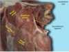
Cow Lateral Pelvis Image

Piriformis m.
(Carnivores Only!!)*
The deep caudal portion of the middle gluteal
- Readily separated
Actions:
- Extend and abduct the hip joint
- Rotate the pelvic limb medially
Innervation:
- Cranial gluteal n.
Blood Supply:
- Cranial gluteal a.
- Lateral circumflex femoral a.
Quadriceps femoris m.
Head 1: Rectus femoris

Only head that crosses the hip and stifle joint
Attachments:
(O): Ilium
(I): Tibal tuberosity
Action:
- Extend the stifle joint
- Flex the hip joint
Innervation:
- Femoral n.
Blood Supply:
- Lateral circumflex femoral a.
- Superficial circumflex iliac a.