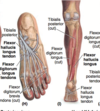
Muscles MD3 Flashcards

1
Q

Deltoid

A

origin: lateral 1/3 of clavicle, acromion, spine of scapula
insertion: deltoid tuberosity
innervation: axillary nn. (C5, C6)

action:

anterior deltoid: humeral flexion & internal rotation

lateral/middle deltoid: humeral abduction

posterior deltoid: humeral extension & external rotation

2
Q

Teres Major

A

origin: posterior surface of inferior angle of scapula
insertion: medial lip of intertubercular/bicipital groove
innervation: lower subscapular nn (C5, C6)
action: adduction & internal rotation of humerus

3
Q

Teres Minor

A

origin: mid part of lateral border of scapula
insertion: inferior facet of greater tubercle of humerus
innervation: axillary nn (C5, C6)

actions:

controls internal rotation of humerus (functional action)

external rotation of humerus

*part of rotator cuff (provide dynamic stability via eccentric contraction for shoulder stabilization/re-enforcement of GH joint capsule)

4
Q

Infraspinatus

A

origin: infraspinous/infraspinatus fossa of scapula
insertion: middle facet of greater tubercle of humerus
innervation: suprascapular nn (C5, C6)

actions:

controls IR of humerus (functional action)

ER of humerus

*part of rotator cuff (provide dynamic stability via eccentric contraction for shoulder stabilization/re-enforcement of GH joint capsule)

5
Q

Supraspinatus

A

origin: supraspinous/supraspinatus fossa of scapula
insertion: superior facet of greater tubercle of humerus
innervation: suprascapular nn (C5, C6)

actions:

prevent superior translation of humeral head during abduction (protect supraspinatus, bursae, etc. from getting pinched by humeral head & acromion) (functional action)

initiate abduction & assist deltoid in abduction of humerus

*part of rotator cuff (provide dynamic stability via eccentric contraction for shoulder stabilization/re-enforcement of GH joint capsule)

6
Q

Subscapularis

A

origin: subscapular fossa of scapula (anterior)
insertion: lesser tubercle of humerus
innervation: upper & lower subscapular nn (C5-C7)

actions:

control ER of humerus (functional action)

IR & adduction of humerus

*part of rotator cuff (provide dynamic stability via eccentric contraction for shoulder stabilization/re-enforcement of GH joint capsule)

7
Q

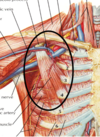
Pectoralis Minor

A

origin: ribs 3-5 (highly variable)
insertion: coracoid process of scapula
innervation: medial pectoral nn (C8, T1)

actions:

stabilizes shoulder, draws scapula anterior & depresses it inferiorly towards the thorax (helps upper extremity reach out forward)

divides axillary aa into its 3 parts

(1st part medial to pec minor, 2nd part posterior, 3rd part lateral)
8
Q

Serratus Anterior

A

origin: lateral aspects of ribs 1-8

insertion:

upper 4 heads- anterior surface of medial border of scapula,

lower 4- inferior angle of scapula

innervation: long thoracic nn (C5-C7)

actions:

protraction of scapula, holds medial border against thoracic wall, upward rotation/external rotation

long thoracic nn can be damaged during mastectomy (winged scapula), unable to raise arm above horizontal

9
Q

Pectoralis Major

A

origin:

clavicular- ant, medial 1/2 of clavicle

sternal- ant sternum, 1st 6 costal cartilages, external oblique aponeurosis

insertion: lateral lip of intertubercular/bicipital groove
innervation: lateral & medial pectoral nn (clavicle C5, C6, sternum C7-T1)

actions:

adduction & IR of humerus, draws scapula anterior & inferior

10
Q

Triceps brachii

A

origin:

long head- infraglenoid tubercle of scapula

lateral head- posterior humerus, superior to spiral groove (radial groove)

medial head- posterior humerus, inferior to spiral groove (radial groove)

insertion: olecranon process of ulna
innervation: radial nn (C6-C8)
actions: extension at elbow

heads recruited additively as needed, medial first (workhorse), lateral (strongest), long (last)

11
Q

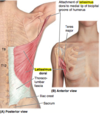
Latissimus Dorsi

A

origin: spinous processes of T7-L5, inferior angle of scapula, thoracolumbar fascia, iliac crest of sacrum, inferior 3 or 4 ribs
insertion: floor of intertubercular/bicipital groove of humerus
innervation: thoracodorsal nn (C6-C8)
actions: adduction, IR & extension of the humerus
12
Q

Biceps brachii

A

brachial aa, musculocutaneous nn (C5, C6)

Long head

origin: supraglenoid tubercle, thru glenoid labrum (constant tension causes slap lesions)

Short head

origin: coracoid process
insertion: radial tuberosity (medial radius) & bicipital aponeurosis
action: primary supinator of forearm, also resistive elbow flexion (hand position dependent)

13
Q

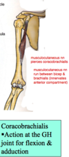
Coracobrachialis

A

brachial aa, musculocutaneous nn (C5-C7)

origin: coracoid process
insertion: mid 1/3 of medial humerus
actions: humeral adduction, flexion @ GH joint (shoulder)
- musculocutaneous nn pierces it
14
Q

Brachialis

A

radial recurrent aa, musculocutaneous nn (C5, C6)

origin: anterior humerus
insertion: coronoid process & tuberosity of ulna
actions: primary elbow flexor (active throughout elbow flexion)
- musculocutaneous nn runs between biceps brachii & brachialis

15
Q

Brachioradialis

A

radial recurrent aa, radial nn (C5, C6)

origin: lateral supracondylar ridge of humerus
insertion: radial styloid process (distal radius)
actions: elbow flexion

only flexor in the posterior compartment, superficial layer

16
Q

Pronator teres

A

ulnar & radial aa, median nn (C6, C7)

origin: medial epicondyle of humerus & coronoid process of ulna
insertion: lateral aspect of mid radius
actions: pronation of forearm, elbow flexion
- superficial anterior compartment

17
Q

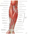
Flexor Carpi Radialis

A

radial aa, median nn (C6, C7)

origin: medial epicondyle of humerus
insertion: base of 2nd & 3rd metacarpals
actions: flexion & abduction of wrist
- superficial anterior compartment
18
Q

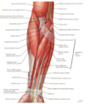
Flexor Carpi Ulnaris

A

ulnar aa, ulnar nn (C8, T1)

origin: medial epicondyle of humerus
insertion: pisiform, hook of hamate, base of 5th metacarpal
actions: flexion and adduction of wrist
- anterior superficial compartment
19
Q

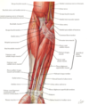
Palmaris Longus

A

ulnar aa, median nn (C7, C8)

origin: medial epicondyle of humerus
insertion: flexor retinaculum & palmar aponeurosis
actions: wrist flexion, tenses palmar aponeurosis
- anterior superficial compartment
20
Q

Abductor Pollicis Longus

A

origin: proximal ulna, radius, interosseus membrane
insertion: base of 1st metacarpal
action: abduction & extension of thumb
innervation: radial nn (deep radial nn) (C7, C8)

*deep layer on posterior forearm

21
Q

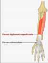
Flexor Digitorum Superficialis

A

origin: anterior oblique line of radius, coronoid process of ulna, medial epicondyle of humerus
insertion: middle phalanges
innervation: median nn (C7, C8, T1)
action: flexion of middle phalanges @ PIP, then flexion of proximal phalanges @ MCP

22
Q

Flexor Digitorum Profundus

A

origin: ulna & interosseus membrane
insertion: distal phalanges (2-5)
innervation: median nn & ulnar nn (4th & 5th digits)
action: flexion of distal phalanges @ DIP

*deep anterior forearm

23
Q

Flexor Pollicis Longus

A

origin: radius
insertion: distal phalanx of thumb
innervation: median nn
action: flexion of thumb

*deep anterior forearm

24
Q

Pronator Quadratus

A

origin: ulna
insertion: radius
innervation: median nn
action: pronation of forearm

*deep anterior forearm

square “quad” shaped muscle

25
Q

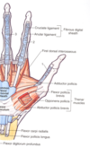
Abductor pollicis brevis

A

origin: flexor retinaculum, scaphoid, trapezium
insertion: lateral proximal phalanx of thumb
innervation: median nn
action: abduction of thumb

*thenar (palmar base of thumb)

-intrinsic muscle

26
Q

Flexor Pollicis Brevis

A

origin: flexor retinaculum, scaphoid, trapezium
insertion: proximal phalanx of thumb
innervation: median nn
action: flexion of thumb @ MCP joint

*thenar, intrinsic

27
Q

Opponens Pollicis

A

origin : flexor retinaculum, scaphoid, trapezium

insertion: lateral 1st metacarpal
innervation: median nn
action: opposition of thumb

*thenar, intrinsic

*deep to flexor and abductor pollicis brevis

28
Q

Adductor Pollicis

A

-ulnar nn.

29
Q

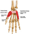
Abductor Digiti Minimi

A

origin: pisiform
insertion: medial 5th proximal phalanx
innervation: ulnar nn
action: abduction of 5th digit, aids in flexion of proximal 5th phalanx

*hypothenar (muscles on medial side of palm, pinky)

30
Q

Flexor Digiti Minimi

A

origin: hook of hamate & flexor retinaculum
insertion: medial 5th proximal phalanx
innervation: ulnar nn.
action: flexion of 5th proximal phalanx

*hypothenar

31
Q

Opponens digiti minimi

A

origin: hook of hamate & flexor retinaculum
insertion: medial border of 5th metacarpal
innervation: ulnar nn.
action: opposition of 5th digit/pinky

*hypothenar
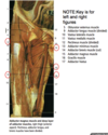
*deep to flexor & abductor digiti minimi

32
Q

Extensor Pollicis Longus

A

origin: posterior middle 1/3 of ulna
insertion: distal phalanx of thumb
innervation: radial nn.
action: extension of thumb (IP, MCP, CMC jts)

*deep posterior forearm

33
Q

Extensor Pollicis Brevis

A

origin: posterior distal 1/3 of radius
insertion: proximal phalanx of thumb
innervation: radial nn.
action: extension of thumb @ MCP & CMC

*deep muscle of forearm (extrinsic)

34
Q

Extensor Carpi Radialis Longus

A

origin: lateral supraepicondylar ridge of humerus
insertion: posterior base of 2nd metacarpal
innervation: radial nn.
action: flexion & abduction of wrist, active during fist clenching

*medial to brachioradialis

35
Q

Extensor Carpi Radialis Brevis

A

origin: lateral epicondyle of humerus (common extensor tendon)
insertion: posterior base of 3rd metacarpal
innervation: radial nn
action: extension & abduction @ wrist

36
Q

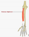
Extensor Digitorum

A

origin: lateral epicondyle of humerus (common extensor tendon)
insertion: distal phalanges 2-5 (via extensor expansions)
innervation: radial nn.
action: extension of wrist & digits 2-5 primarily @ MCP joint

37
Q

Extensor Indicis

A

origin: posterior distal 1/3 of ulna
insertion: extensor hood of 2nd digit
innervation: radial nn.
action: independent extension of 2nd digit

*deep posterior forearm

38
Q

Supinator

A

origin: lateral epicondyle of humerus, ulna, radial collateral & annular ligaments
insertion: lateral, posterior & anterior proximal radius
innervation: radial nn.
action: supination of forearm

*deep posterior forearm

39
Q

Lumbricals

A

origin:

lateral 2: lateral side of FDP tendons of 2 & 3rd digits

medial 2: FDP tendons of 3-5th digits

insertion: lateral extensor expansions

innervation:

lateral 2: median nn.

medial 2: ulnar nn.

action: flexion @ MCP, extension @ IP joints

40
Q

Interosseus Muscles

A

DAB (dorsal) & PAD (palmar)

innervation: ulnar nn.

41
Q

Extensor Hood

A

tendonous aponeurosis of 2-5th digits flatten and wrap around distal metacarpals & proximal phalanges forming a dorsal hood that holds extensor tendons in place

42
Q

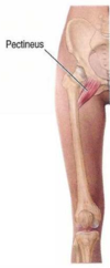
Pectineus

A

O/I: superior pubic ramus -> pectineal line of humerus

innervation: Femoral nn.

Actions: adduction & flexion of thigh, internal rotation of humerus

43
Q

Iliopsoas

A

psoas major + iliacus (join @ midpoint)

O/I: lumbar vertebrae, iliac crest-> lesser trochanter of femur

innervation: lumbar rami (L1-3), femoral nn.
actions: flexion of thigh, trunk flexion when legs grounded, maintains lordosis of lumbar spine (tightness of muscle = incr lordosis)

44
Q

Sartorius

A

longest muscle in body, spans two joints (hip & knee)

O/I: anterior superior iliac spine-
> pes anserine

innervation: femoral nn.
actions: “tailor’s muscle”, flexion of thigh/hip, flexion of knee, weak ext rotation of thigh, weak abduction of thigh, primarily synergistic

45
Q

Quadriceps Femoris

A

Rectus Femoris:

O: anterior inferior iliac spine

-spans two joints (hip & knee), longest muscle in quadriceps femoris, “kicking muscle”

Vastus Lateralis:

O: greater trochanter of femur, lateral lip of linea aspera

Vastus Intermedius:

O: anterolateral shaft of femur

Vastus Medialis:

O: intertrochanteric line & medial lip of linea aspera of femur

All come together to insert on quadriceps tendon-> patella-> patellar ligament to attach onto tibial tuberosity

Should be 3:2 stronger than hamstring, commonly 3 x stronger

Actions: flexion of hip/thigh (only rectus femoris), extension of knee (all)

Innervation: femoral nn.

46
Q

Adductor Brevis, Adductor Longus, Adductor Magnus

A

O/I: inferior ischial & pubic ramus-> linea aspera & adductor tubercle of femur (adductor magnus, hamstring part)

innervation: obturator nn. (all), magnus = obturator nn. (adductor part), tibial nn. (hamstring part)
actions: adduct thigh (brevis, longus, adductor part of magnus), may flex thigh (brevis), ext thigh (hamstring part of magnus)

*obturator nn splits ant/post @ brevis

magnus is most powerful adductor

adductor hiatus (magnus) allows transmission of femoral aa & vv from adductor canal to popliteal fossa

47
Q

Gracilis

A

O/I: inferior pubic ramus-> pes anserine

innervation: obturator nn.
actions: adduction of thigh, flexion of knee, internal rotation of humerus when knee flexed

48
Q

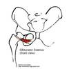
Obturator Externus

A

O/I: obturator membrane in obturator foramen-> trochanteric fossa on greater trochanter of humerus

innervation: obturator nn.
actions: external rotation of thigh, stabilizes femoral head in acetabulum

49
Q

Gluteus maximus

A

-largest and most superficial (superficial gluteal, primary movers of hip)

O/I; iliac crest, sacrum, coccyx -> iliotibial (IT) band, some fibers to gluteal tuberosity of femur

innervation: inferior gluteal nn
actions: mainly hip extension (esp. up inclines) & external rotation of femur

50
Q

Gluteus Medius & Minimus

A

O/I: gluteal surface of ilium (beneath iliac crest, deep to gluteus maximus)-> greater trochanter of femur

innervation: superior gluteal nn.
actions: abduction of thigh, internal rotation of femur, medius stabilizes pelvis during gait

Trendelenburg sign/gait: weakness of these muscles

51
Q

Tensor Fasciae Latae

A

O/I: anterior superior iliac spine -> iliotibial band

innervation: superior gluteal nn.
actions: flexion @ hip, abduction and internal rotation of femur
- fusiform muscle (wide in middle, narrow at ends)

52
Q

Piriformis

A

O/I: Sacrum -> greater trochanter of femur

innervation: anterior rami of S1 & S2
actions: external rotation of extended thigh, abductor of flexed thigh
- superior gluteal nn & vessels travel above piriformis
- inferior gluteal nn. & vessels travel below piriformis, along w/ sciatic nn.
- piriformis syndrome (sciatica)- irritation of sciatic nn by piriformis causes pain in butt radiating down leg (sciatic nn.)

53
Q

Triceps Coxae

A

Obturator Internus:

O/I: ischiopubic ramus & obturator membrane -> medial part of greater trochanter of femur

innervation: nn to obturator internus (L5, S1)
actions: external rotation of extnded thigh, abduction of flexed thigh

*located b/w superior & inferior gemelli

Gemelli:

O/I: ischial spine (superior) & ischial tuberosity (inferior) -> medial part of greater trochanter of femur (all 3 tendons of triceps coxae join and insert onto the medial greater trochanter)

innervation: nn to obturator internus (superior), nn. to quadratus femoris (inferior) (L5, S1)
actions: external rotation of thigh

54
Q

Quadratus Femoris

A

O/I: ischial tuberosity -> intertrochanteric crest

innervation: nn. to quadratus femoris (L5, S1)
actions: strong external rotation of thigh
- located inferior to inferior gemellus

55
Q

Semitendinosus, Semimembranosus (part of hamstring)

A

O/I: ischial tuberosity -> pes anserine (semitendinosus), posterior medial condyle of tibia, popliteal fossa, popliteal ligament (semimembranosus)

innervation: tibial branch of sciatic nn.
actions: extension of hip, flexion of knee, internal rotation w/ flexed knee
- commonly used in ACL repair (semitendinosus)

56
Q

Biceps Femoris (workhorse of the hamstring)

A
O/I:
ischial tuberosity (long head), linea aspera & lateral supracondylar line of femur (short head) -\> fibular head, LCL of knee

innervation: tibial branch of sciatic (long head), common peroneal branch of sciatic (short head)
actions: hip extension, knee flexion, external rotation of flexed knee

*hamstring strains are twice as likely as quads, commonly d/t co-contracture (e.g. running), avulsion at ischial tuberosity

57
Q

Pes Anserine

A
  • insertion of tendons from Sartorius, Gracilis, SemiTendinosus (Say Grace SomeTimes) onto anteriomedial upper tibia
  • looks like a goose’s foot
  • common site of tendinitis/bursitis
58
Q

Tibialis Anterior

A

O/I: upper 1/3 & lateral condyle of tibia -> medial cuneiform & base of 1st metatarsal

innervation: deep peroneal nn (branch off common peroneal)
actions: dorsiflexion, inversion of ankle
- on lateral surface of tibia, most superficial, strongest dorsiflexor

59
Q

Extensor Digitorum Longus

A

O/I: lateral condyle of tibia & anterior shaft of fibula-> middle and distal phalanges

innervation: deep peroneal nn.
actions: extension of 4 digits (not hallicus), dorsiflexion
- most lateral anterior leg muscle, lies on medial side of fibula

60
Q

Extensor Hallucis Longus

A

O/I: anterior interosseus membrane-> distal 1st phalanx

innervation: deep peroneal nn.
actions: extension of great toe, dorsiflexion
- deep muscle, lies between EDL & tibialis anterior

61
Q

Peroneus (Fibularis) Tertius

A

O/I: distal anterior fibula-> 5th metatarsal

innervation: deep peroneal nn.
actions: aids in dorsiflexion, eversion of foot, ankle proprioception

62
Q

Peroneus Longus

A

O/I: upper lateral fibula-> 1st metatarsal & medial cuneiform

innervation: superficial peroneal nn.
actions: plantar flexion & eversion of foot
- posterior to lateral malleolus, underneath foot, thru groove in cuboid
- antagonistic to peroneus tertius (plantar flexion) and agonistic (eversion)
- longer, more superficial, originates higher up on tibia vs brevis

63
Q

Peroneus Brevis

A

O/I: lower lateral fibula-> lateral base of 5th metatarsal

innervation: superficial peroneal nn.
actions: eversion of foot, plantar flexion
- deep to peroneus longus, runs posterior to lateral malleolus
- common avulsion site (Jones fx)

64
Q

Gastrocnemius

A

O/I: posterior condyles of femur-> achilles tendon

innervation: tibial nn.
actions: plantar flexion (propulsion phase of walking), knee flexion
- medial & lateral heads, fast-twitch muscle

65
Q

Soleus

A
  • deep to gastrocnemius, joins it distally to become achilles tendon (most powerful tendon)
  • workhorse of plantar flexion, (active in standing and postural balance)
  • popliteal aa & tibial nn lay underneath it
    innervation: tibial nn
66
Q

Plantaris

A

O/I: lateral supracondylar ridge of femur-> achilles

innervation: tibial nn
actions: weak plantar flexion, may be more significant in proprioception
- absent in 5-10%, used for grafting to hand tendons

67
Q

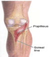
Popliteus

A

O/I: lateral femoral condyle-> proximal posterior tibia

innervation: tibial nn.
actions: knee flexion, opposite screw home mechanism (turns femur laterally)
- inferior floor of popliteal fossa

68
Q

Flexor Hallucis Longus

A

O/I: posterior mid fibula-> distal 1st phalanx

innervation: tibial nn.
actions: powerful great toe flexor, supports medial longitudinal arch of foot
- runs under flexor digitorum longus tendon under navicular bone

69
Q

Flexor Digitorum Longus

A

O/I: posterior tibia-> distal lateral 4 phalanges

innervation: tibial nn.
actions: flexion of phalanges, plantar flexion, supports longitudinal arches
- passes over tibialis posterior @ distal tibia

70
Q

Posterior Tibialis

A

deepest/most anterior muscle in posterior compartment

innervation: tibial nn.
actions: works w/ tibialis anterior to invert foot, supports medial longitudinal arch
- inserts on navicular and cuneiform bones
- b/w flexor digitorum longus & flexor hallucis longus

71
Q
A
72
Q

1st layer of plantar muscles

A

Abductor Hallucis

O/I: calcaneus-> medial 1st proximal phalanx

innervation: medial plantar nn.
actions: abduction & flexion of great toe

Flexor Digitorum Brevis

O/I: calcaneus-> both sides of middle phalanges 2-5

innervation: medial plantar nn.
actions: flexion of digits 2-5

Abductor Digiti Minimi

O/I: calcaneus-> lateral 5th proximal phalanx

innervation: lateral plantar nn.
actions: abduction & flexion of 5th digit

73
Q

2nd layer of plantar muscles

A

Quadratus Plantae

O/I: calcaneus-> posterolateral flexor digitorum longus tendon

innervation: lateral plantar nn
actions: assists FDL in flexion of digits 2-5, keeps FDL stable when lumbricals pull on it during flexion

Lumbricals

O/I: FDL tendon to medial aspect of digits 2-5

innervation: lumbrical #1 = medial plantar nn, #2-4 = lateral plantar nn
actions: flexion of proximal phalanges, extension of middle & distal phalanges of digits 2-5

74
Q

3rd layer of plantar muscles

A

Flexor Hallucis Brevis

O/I: cuboid & lateral cuneiform-> base of 1st proximal phalanx

innervation: medial plantar nn
actions: flexion of great toe

Adductor Hallucis (oblique & transverse heads)

O/I: Plantar ligaments of MTP joints (oblique), base of metatarsals 2-4 (transverse)-> lateral aspect of 1st proximal phalanx

innervation: lateral plantar nn.
actions: stabilizes transverse arch by bringin metatarsals medially

Flexor Digiti Minimi

O/I: base of 5th metatarsal-> proximal 5th phalanx

innervation: lateral plantar nn.
actions: flexion of 5th proximal phalanx

75
Q

4th layer of plantar muscles

A

Plantar Interossei (3 muscles, PAD)

innervation: lateral plantar nn.
actions: adduction of digits 2-4

Dorsal Interossei (4 muscles, DAB)

innervation: lateral plantar nn.
actions: abduction of digits 2-4

*both involved in flexion @ MTP

*2nd toe is midline in the foot (cannot abduct)

76
Q

Extensor Digitorum Brevis, Extensor Hallucis Brevis (dorsal foot muscles)

A

innervation: deep peroneal nn.
actions: aids ‘long’ counterparts in extension of digits 2-4 (EDB), 1st toe (EHB)

77
Q

SGT FOS

A

Sartorius- Femoral nn

Gracilis- Obturator nn

semiTendinosis- Sciatic nn