Midterm - external eye exam conditions images Flashcards
(37 cards)
Corneal scarring
Grayish white opacity from old injury or keratitis (inflammation of cornea). Cloudiness may overlap pupil edges

Kaiser-Fleisher Ring
golden to red-brown ring in periphery of cornea. Caused by copper deposition, suggests Wilson Disease

Corneal abrasion
Painful on initial inspection. Can mimic infection, but history will have foreign object or trauma.

Corneal infection
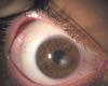
Corneal Ulcer (infectious keratitis)
Caused by bacteria, fungal, parasitic.
dendritic lesions = herpes simplex keratitis

Acute iritis/ anterior uveitis

Synechia from acute iritis/ anterior uveitis
affected pupil is smaller and will NOT dilate

Acute angle closure glaucoma/ narrow angle glaucoma
sudden increase in intraocular pressure happens when aqueous humor drainage is blocked.
symptoms: acute onset with severe, deep, aching pain in the eye (unilateral). Redness, blurry vision, dilated pupils, headache, nausea, vomiting.

Coloboma
absence or defect of iris tissue

Prolapsed iris

Adie’s tonic pupil
Due to parasympathetic neve degeneration. May have blurry vision and photophobia

(Right) CN 3 palsy
ptosis

Horner Syndrome
miosis, ptosis, [facial] anhidrosis

Argyll-Robertson pupils
bilaterally small, irregular shaped pupils.
do not react to light, do accommodate

Dacryoadenitis
lacrimal gland

Dacryocystitis
puncta side of eye, painful

Acute dacryocystitis

Ectropion

Entropion

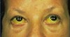
Blepharitis
red inflammed lid margins. NOT turning. Greasy, crusted with scaling debris on lashes.

Anterior blepharitis (due to seborrheic dermatitis)

Severe anterior blepharitis (due to staphylococcal infection)

External hordeolum (stye)

Internal hordeolum