Macula and Choroid 2 Flashcards
(30 cards)
What is Cystoid Macular Oedema?
Macular Oedema as a result of fluid accumulation (cysts) at the Outer Plexiform Layer & Inner Nuclear Layer of the fovea which is due to a breakdown of the blood-retinal barrier.
Is acute/short term cystoid macula oedema serious?
It may not be serious as it can improve without treatment
Is chronic Cystoid Macula Oedema (CMO) serious and if so why?
Yes it is serious as it may cause a permanent decline in central vision.
It is also serious as it can be difficult to treat.
Cysts may coalese to form a lamellar hole.
What are the clinical signs of Cystoid Macula Oedema (CMO)?
Clinical signs:
- Loss of foveal depression – i.e. thst the macula swells or doesn’t dip in
- Cysts may be visible on OCT – appear as hyporeflective spaces within the retina ( this means they look like dark spots)
- FA typical “flower-petal”pattern of late hyperfluorescence –due to dye accumulation with time in the Outer Plexiform Layer cysts

What are the possibel causes of Cystoid Macula Oedema?
- Post surgery
- Diabetes
- Wet AMD
- Retinal vein occlusion
- Uveitis
- Epiretinal membranes
- Radiation retinopathy
- Choroidal tumours
- Retinitis pigmentosa
- Prostaglandin analogue eyedrops for glaucoma
- Macula telangectasia
- Coats disease
- And others…
What is Irvine gass syndrome and what is it also known as?
Pseudophakic cystoid macular edema (CME), also known as Irvine-Gass syndrome, is one of the most common causes of visual loss after cataract surgery.
Basically it is the term given to a CMO developing after cataract surgery.
What is the main symptom of Cystoid Macula Oedema?
Painless loss of central vision ( this can vary from 6/12 to 6/60)
True or false - After a diabetic’s cataract surgery CMO is more likely to occur especially if they have diabetic retinopathy to begin with.
True
•In diabetes, post-cataract surgery CMO occurs in 32% without DR, and 81% of diabetics with DR (Diabetic retinopathy)
–Pre-existing diabetic macular oedema should be treated before cataract surgery whenever possible
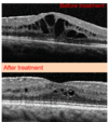
What is the following image showing?

CMO ( after cataract surgery)
Why do px require a follow up appt 6-12 weeks after cataract surgery?
To ensure CMO is not developing.
What is the treatment for Irvine Gass Syndrome?
- Topical Non-steroidal anti-inflammatory drugs (NSAIDs)
- Topical corticosteroids
- Anti-VEGF?
[After treatment you can see that the CMO has settled but you do have these slight spaces, intraretinal remnants of the cystic spaces]
What does pathological/degenerative myopia cause?
It causes degernative changes in the sclera, Choroid and RPE.
What prescription and axial length is pathological/degenrative myopia associated with?
A prescription above -6.00D
and an axial length greater than 26mm.
What is the prevalence of pathological myopia?
It affects 2-10% of the population and is race dependant
What are some examples of systemic associations of pathological/degenerative myopia?
- Down syndrome
- Marfan syndrome
- Prematurity
- Ehlers-Danlos syndrome
How may a myopic fundus typically present?
•As a Pale tessellated (tigroid) fundus
–(Diffuse reduction of RPE with increased visibility of choroidal vasculature
•
•Focal chorio-retinal atrophy –patchy areas where choroid (and possibly) sclera become visible
•Anomalous optic nerve head –It may have a Small, large or tilted appearance
•Peripapillary chorio-retinal atrophy –This is very Common
–This is atrophy surrounding the nerve head, it also includes Peripapillary intrachoroidal cavitation which is the presence of a small yellow-orange area inferior to disc ( this can cause a visual field defect)
•Can present with an Acquired optic disc pit –Due to expansion of peripapillary area over time i.e. with growth and elongation a pit within the optic disc may form.
•Can present with Lacquer cracks
•Can present with Subretinal haemorrhages (may develop from lacquer cracks)
•Can present with Choroidal neovascularisation
•Can present with Fuch’s spot
Peripapillary intrachoroidal cavitation can cause a visual field defect that is mistaken for what condition?
Glaucoma
What are lacquer cracks and how are they seen on a fundus?
Why do lacquer cracks form?

How common is Choroidal neovascularisation in pathological myopia?
•CNV in ~10% of highly myopic eyes
In pathological myopia does CNV cause maximal sensory retinal elevation or minimal sensory retinal elevation and why is this an important thing to note?
It causes minimal sensory retinal elevation - this is an important thing to note when comparing to a condition such as wet AMD, in which CNV is likely to cause a much more significant sensory retinal elevation I( i.e. it basically helps with diffrentiating between our diagnosis)
In what pathologically myopic patients in CNV likely to be a symptom?
px over 40-50
True or false - myopic eyes with CNV tend to have a worse prognosis than those with wet AMD?
False - myopic eyes with CNV have a better prognosis than px with CNV who have wet AMD
What does it mean to be sub-foveal?
To be just below the fovea
What does it mean to be Juxtafoveal?
Next to the fovea

