Lymphadenopathies & Lymphomas - Harrington Flashcards
Describe the means by which lymphocytes enter and exit lymphoid organs such as lymph nodes.
Enter the lymph node from the blood via HEVs. Exit via the efferent lymphatics after clonal expansion.
How does the setting of clonal expansion differ between B and T cells?
B cells migrate to the cortex (mantle zone) and expand in the germinal center, then migrate to the marginal zone to differentiate into plasma cells.
T cells migrate to the paracortex and expand there (presumably following costimulation by B cells).
A patient experiences enlargement of “glands” around her cervical spine. They are painful, and biopsy reveals secondary lymphoid follicles. What can you say about the etiology of this condition?
Lymphoid architecture is maintained and the LAD is painful; This is a reactive lymphadenitis, probably relating to an infection.
What criteria on clinical or histological exam can help distinguish a reactive from a neoplastic lymphadenopathy?
Clinical: Duration, size, location, extent, tenderness, mobility.
Histologic: Cellular architecture, dominant cell type, atypias, flow cytometry (immunophenotype)
What are some possible causes of reactive lymphadenopathy?
Describe 4-5 patterns of reactive lymphadenopathy.
Infectious, autoimmune, drugs, foreign body, sarcoidosis, and many other eponymous conditions…
**Follicular/Paracortical hyperplasias, Sinus histiocytosis, **Necrotizing and Granulomatous.
Give the histologic patterns found in lymph nodes under the following conditions:
Viral infection
Rheumatoid arthritis
Tumors in area of drainage
Cat scratch fever (Bartonella henselae)
Toxoplasmosis
Viral: Paracortical hyperplasia
RA: Follicular hyperplasia
Tumors: Sinus histiocytosis
Cat scratch: Necrotizing
Toxoplasmosis: Follicular hyperplasia
(from optional webcast)
What fraction of lymphomas are B-cell?
What fraction of NHLs are nodal?
About 80% of lymphomas are B-cell related.
2/3 of NHLs are nodal.
(from optional webcast)
Describe the Ann Arbor clinical staging system for lymphoma.
What other staging system exists
Stage I (involves 1 LN), II (2 LNs on same side of diaphragm), III (involvement on both sides of diaphgragm), IV (extralymphatic infiltration). A/B suffix (B = presence of B symptoms)
International Prognostic Index (IPI)
(from optional webcast)
A precise etiology for NHLs is not known. However, there are several conditions or pathophysiologies that are thought to contribute. Name 3.
- Chronic inflammation (increases the chance of developing a mutant clone)
- Translocations (usually involving IgH locus)
- Accidents in normal clonal rearrangement
Differentiate between low-grade and high-grade lymphomas with respect to their cell size, localization, and survival.
Low grade: Indolent, small cells, disseminated, kills slowly but surely (incurable; short-term survival is good but long-term is bad)
High grade: Aggressive, large cells, localized, kills quickly but plateaus (curable; long-term survival exceeds low-grades)
What are the most common lymphomas?
DLBCL (Diffuse Large B-Cell Lymphoma)
Follicular Lymphoma
Describe the structure of rituximab.
What is its mechanism of action?
Rituximab is a chimerized IgG (murine Fab, human Fb).
Targets CD20 antigen to eliminate B cells via CDC, ADCC, activation of apoptosis, and delivery of ionizing payload (?)
Follicular Lymphoma
How common is it?
Who is the “sterotypical patient”?
How does it present cilnically?
Follicular Lymphoma
Fairly common; 20% of all lymphomas.
An older (60yo) male.
Generalized, painless adenopathy.
Follicular Lymphoma
What is the most common cytogenetic aberration?
How is its outlook?
Follicular Lymphoma
t(14;18)
Generally indolent; 40% transform to aggressive lymphomas (DLBCL, Burkitt). Advanced stages previously incurable (Anti-CD20 being the first therapy to reduce mortality).
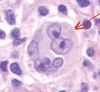
What are reed-sternberg cells?
What markers do they have?
What disease do they indicate?
Multinucleate, B cell derivatives
CD15 and CD30
Hodgkin’s Lymphoma
What are some differences between Hodgkin’s and Non-Hodgkin’s lymphoma?
Hodgkin’s Lymphoma
- Central, Axial Nodes
- Spread contiguously
- Bone Marrow Involvement Rare
Non-Hodgkin’s Lymphoma
- Peripheral Lymph nodes
- Spread non-contiguously
- Bone Marrow Involvement Frequent
What are the 4 classical types of Hosdgkin’s Lymphoma?
What is the non-classical type?
Nodular Sclerosis
Mixed Cellularity
Lymphocyte Predominant
Lymphocyte depleted
Nodular Lymphocye Predominant
Nodular Sclerosis Hodgkin’s Lymphoma
What age is more affected?
Which Gender is more affected?
Site?
Cell Markers?
Morphology?
15-35 years old
M=F
Mediastinal Nodes
CD15, CD30
Lacunar Cells, Dense sclerosis
Mixed Cellularity Hodgkin’s Lymphoma
What age is more affected?
Which Gender is more affected?
Site?
Cell Markers?
Morphology?
Bimodal Ages (young and Old)
M>F
Cervical, Axillary
CD15, CD30
Inflammatory milieu
Nodular Lymphocyte Predominant
What age is more affected?
Which Gender is more affected?
Site?
Cell Markers?
Morphology?
30-50 years old
M>F
Cervical, Axillary
CD20, CD45 (Remember this is the abnormal HL)
Popcorn Cells
Ebstein Barr Virus
What family of viruses does it belong to?
What is it’s genome?
Is it enveloped?
Herpes Virus (specifically gammaherpes)
Double-stranded DNA
Yes
What happens with EBV during lytic infection, starting from infection?
1) EBV binds to and fuses with cell
2) Viral genome goes to nucleus
3) VHS chews up host mRNA while VP16 starts viral transcription
4) EBV alpha genes are transcribed
5) EBV beta genes are transcribed
6) EBV gamma genes are transcribed
7) Viral DNA is packaged into capsids
8) Capsids bud off through cell membrane
How does EBV manage to stay latent in a healthy host?
- double stranded DNA is able to integrate into genome
- Viral genome is repressed by methylated histones
- EBNA1 maintains histone repression
- Once B cells turns into Plasma cell, EBNA1 activates viral genes
What are EBERs?
Ebstein Barr Encoded RNAs
RNA produced in all infected cells which is never transcribed
When is EBV acquired?
How does the disease change with age of infection?
The majority are infected in early childhood, with virtually everyone infected by young adulthood.
In young children, it causes a mild cold; in older children/adults, in causes infectious mononucleosis
What characterizes infectious mononucleosis?
How do we test for it?
Expansion of CD8+ T cells (Downey Cells) and B cells. Massive production of both autoimmune and xenogenic antibodies.
We test for the heterophile antibodies produced.
How does EBV cause cancer?
4 mechanisms
Direct Transformation of Cells
Cell Mutations
Growth factor Storm
Chronic Inflammation
Does EBV cause cancer through direct transformation of cells?
Unclear; it is associated with increased risks of cancer and viral proteins have been associated with cancer in animal models . However, most of the population is positive and EBV caused cancer is still relatively rare.
classify the following according to expected cellular architecture (follicular/nodular or diffuse)
- Burkitt lymphoma
- Mantle cell lymphoma
- CLL/SLL
- Follicular lymphoma
- Diffuse large B-cell lymphoma
- Marginal zone lymphoma
- diffuse
- nodular/follicular
- diffuse
- nodular/follicular
classify the following according to expected tumor size (small, medium/large)
- CLL/SLL
- lymphodysplastic lymphoma
- follicular lymphoma
- diffuse B-cell lymphoma
- mantle cell lymphoma
- marginal zone lymphoma
- burkitt lymphoma
- small
- small
- small
- medium/large
- any size
- small
- medium/large
As a general rule, which (among B-NHLs) is more aggressive?
- small cells vs. med/large cells
- nodular/follicular vs. diffuse
- med/large
- diffuse
med/large and diffuse includes Diffuse B-cell Lymphoma and Burkitt Lymphoma
Name the cytogenetic translocation associated with each
- Follicular lymphoma
- Burkitt lymphoma
- Mantle cell lymphoma
- t(14;18)
- t(8;14)
- t(11;14)
Among small B-NHLs, identify the disease based on the following immunophenotypes:
- CD5(-), CD10(+)
- CD5(+), CD23(-)
- CD5(+), CD23(+)
- CD5(-), CD10(-)
- Follicular lymphoma
- Mantle cell lymphoma
- CLL/SLL
- Marginal zone lymphoma
Give the major cytogenetic translocation associated with Burkitt lympoma. Name (2) others.
- major: t(8;14)
- minor: t(2;8) and t(8;22)
What is the classic description of LM findings in Burkitt lymphoma?
Explain what’s happening here
“Starry sky”
The ‘sky’ backdrop is tumor cells. The ‘stars’ are tingible macrophages. The growth of tumor cells is so rapid that they effectively starve/crowd each other out, leading to an accumulation of dead tumor cells. The tingible macrophages are attempting to clear out the debris from these dead cells.
Match the following cytogenetic translocaitons to its associated B-NHL:
- t(11;14)
- t(2;8)
- t(11;18)
- t(8;22)
- t(8;14)
- t(14;18)
- Mantle cell lymphoma
- Burkitt lymphoma
- Marginal zone lymphoma
- Burkitt lymphoma
- Burkitt lymphoma
- Follicular lymphoma
Identify: CD5(+) with cyclin D1 overexpression
Mantle cell lymphoma
Identify: c-MYC and BCL2 overexpression
Follicular lymphoma
Identify: CD30(+) and CD15(+)
Classic Hodgkin Lymphoma
Identify

Burkitt lymphoma (“starry sky”)
Identify the cell indicated by the red arrow

Reed-Sternberg cell (Hodgkin Lymphoma)
What is the cytogenetic translocation observed in follicular lymphoma? Explain how this translocation results in disease.
t(14;18)
This translocation results in a IgH-BCL2 fusion (IgH heavy chain is on chromosome 14), leading to overexpression of BCL2.
BCL2 prevents apoptosis in cells. Overexpression of this gene blocks normal B-cell apoptosis, leading to an accumulation of B-cells in the lymph node follicles (hence “follicular”)
What is the cytogenetic translocation observed in mantle cell lymphoma? Explain how this translocation results in disease.
t(11:14)
This translocation results in a CyclinD1-IgH fusion, leading to overexpression of Cyclin D1. Cyclin D1 promotes progression of the cell cycle, leading to proliferation of neoplastic B-cells.
What is the cytogenetic translocation usually observed in Burkitt lymphoma? Explain how this translocation results in disease.
t(8;14)
This translocaiton results in a c-MYC-IgH fusion, leading to overexpression of c-MYC. c-MYC is a transcription factor - its overexpression leads to overproduction of growth factors and increased growth/proliferation of neoplastic B-cells.
Classify the following as generally “indolent” or “high grade”
- CLL/SLL
- Marginal zone lymphoma
- Burkitt lymphoma
- Lymphoplasmacytic lymphoma
- Diffuse large B-cell lymphoma
- Follicular lymphoma
- Mantle cell lymphoma
- indolent
- indolent
- high grade
- indolent
- high grade
- indolent
- intermediate (has features of both categories)
Where can extranodal marginal zone lymphoma be found?
Mainly MALT tissues (gastric, thyroid, lung, etc)
Is diffuse B-cell lymphoma typically nodal or extranodal?
Both nodal **and **extranodal
Are T/NK cell lymphomas generally indolent or aggressive?
Name (3)
Which is most common in children?
Which is most common in adults?
aggressive
Peripheral T-cell lymphoma, Anaplastic T-cell lymphoma (ALK+), Extranodal NK/T-cell lymphoma
Children: Anaplastic T-cell lymphoma
Adults: Peripheral T-cell lymphoma
Name the most common cell type underlying the following lymphomas:
- Peripheral T-cell lymphoma
- Anaplastic T-cell lymphoma
- Extranodal T-cell lymphoma
- Helper T-cell (Th), rarely cytotoxic
- Cytotoxic T-cell (CTL)
- NK-cell, rarely CTL
Which neoplasm of mature T-cells or NK cells is typically associated with EBV?
Name the anatomical site usually involved in this disease
What geographic regions are most typically associated with this disease?
Extranodal NK/T-cell lymphoma
Sinonasal
Most common in Asia and South America
Identify: CD20(+), CD45(+), “popcorn cells”, B-cell origin
Nodular lymphocyte predominate Hodgkin Lymphoma (NLPHL)
Identify the lymphocytic neoplasm typically associated with ALK gene rearrangements.
What is the gene translocation most commonly associated with ALK in this disease?
In what patient population does this typically present?
Describe the basic clinical features
Anaplastic large-cell lymphoma
t(2;5)
Children and young adults
Usually presents with lymph node and soft-tissue disease
Name (4) clinical variants of Burkitt lymphoma
Sporatic (ileocecal mass)
endemic (EBV, jaw, breast, ovary masses)
Immunosuppression-related (EBV)
Transformation from follicular lymphoma