Lecture 6- Embryology part I Flashcards
primitive atrium
forms the atria
Achievements of early embryonic development
- First two weeks created tissues of the future embryo and future placenta
- The third week created the three germ layers
- Ectoderm, mesoderm and endoderm – primordia of all tissue
- Fourth week created recognisable body form and the mesoderm begins to organise
when does the CVS begin development
10th day after fertilisation- first functional organ to develop
when can the heart be heard
- Can be heard beating on a sonography by week 6
outline the key 5 stages of embryonic development of the CVS
(1) Formation of the primitive heart tube
(2) Cardiac looping
(3) Development of the atria
(4) Formation of the Great vessels
(5) Septation
At the start of this development the CVS system exists as two regions
near the cranial (head end) end of the embryo- cardiogenic fields (derived from the mesoderm)
Cardiogenic fields consist
consists of blood islands which are primitive tissue and mark the beginning of blood, vessel and heart development
Blood islands develop further and
fuse to form two tubes which are called endocardial tubes – one on each side of the embryo

how are the two endocardail tubes brough together
Folding
In the 4th week the embryo begins to folding which puts the heart tissue in the correct position to form the primitive heart tube surrounded by the pericardial sac
how many ways does folding occur
2
- Cephalo-caudal folding
- Lateral folding
Cephalo-caudal folding
Brings cardiogenic filed from cranial entre towards the centre of the embryo to sit in the thoracic region where the heart will be

Lateral folding
- Fuses the two lateral sides of the embryo
- Brings two cardiogenic fields into the midline so they can fuse and form the primitive heart tube

what day is the ptimitive ehart tube form
day 25
characteristics of the primitive heart tube
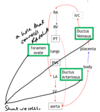
- 6 parts
- no valves
- no barriers between structure
name the 6 parts of the primitive heart tube
- Aortic roots- forms arteries of the aortic arch
-
Truncus arteriosus- outflow of blood
- Involved in the formation of the pulmonary trunk and aorta
- Bulbus cordis- Involved in the formation of the pulmonary trunk and aorta
- Primitive ventricle- forms ventricles
- Primitive atrium- forms the atria
- Sinus venosus- forms part of the right atrium and vena cave

aortic roots
formed at the top of the primitive heart tube
- forms arteries of the aortic arch
Truncus arteriosus
outflow of blood
involved in the formation of the pulmonary trunk and aorta
bulbus cordis
- Involved in the formation of the pulmonary trunk and aorta
primitive ventricles
forms ventricles
sinus venosis
acronymn to learn parts of the primitive heart tube
All The Best Vaccums Are Silver
what happens after the primitive heart tube is formed
(2) Cardiac looping
why must the heart loop
The newly formed heart tube is surrounded by the pericardial sac- as the heart tube grows and elongates, it gets too long for the sac. This means that to fit, it must loop
outline cardiac looping
- The primitive ventricle moves ventrally (coming forward) and to the right
- The primitive node moves dorsally (behind) and to the left
- This puts the inflow portion of the heart (veins and atria) behind the outflow portion (ventricles and arteries) the same shape and orientation as mature hearts


