LE joints and arches Flashcards
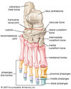
Portions of foot
Hindfoot: talus and calcaneus
Midfoot: navicular, cuboid, cuneiforms (3)
Forefoot: metatarsals, phalanges

Subtalar Joint
- Articulation between the 3 separate inferior articulating surfaces on the talus WITH 3 separate articulating surfaces on the superior aspect of calcaneus that help with articulation
- Function of subtalar jt:
- Translates the motion of the tibia to the foot and vice versa-(takes brunt of force)
- Dampens the rotational forces while maintaining contact with the ground
- Allows for smooth walking over uneven surfaces; pivoting
•Very stable jt - strong ligamentous support includes:
–talocalcaneal ligaments, medial and lateral collateral ligaments
–Described as both uniaxial or multiaxial depending on the reference
•Three separate articulations results in:
–triplanar movement (cuts through 3 cardinal planes) around a single oblique joint axis, moving thru those 3 planes of motion
–Unique jt bc cuts thru the 3 cardinal planes, doesn’t just move within 1
- Subtalar Axis - ~42○ up from the horizontal plane (medial view) and ~ 25○ in from the sagittal plane (lateral view)
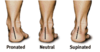
- Motion available: (composite motion) supination (inversion, PF, add) and pronation(eversion, DF, abd)

3 articulations between talus and calcaneus
- anterior talar articular surface
- middle talar articular surface
- posterior talar articular surface

Motions at subtalar jt
•Supination and Pronation are composite motions
*In a Non Weight-Bearing Position (open kinematic chain)
- Supination…a combination of…clacaneal inversion, add, PF
- see big toe
- Pronation ….a combination of….calcaneal eversion, abd, DF
- see lesser toes or no toes
Subtalar jt ligaments
(pic= interosseous talocalcaneal ligs)

- Lateral talocalcaneal ligament
- Dorsal talonavicular ligament
- Interosseous talocalcaneal ligament at the sinus tarsi canal (group of ligs not just one)
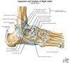
Posterior Talocalcaneal ligament
Tarsal sinus
Space between talus and calcaneus

Cuboid Bone
- Cube like shape
- Most lateral tarsal bone in the distal row of tarsus
- Note cuboid tuberosity on plantar surface-nothing attaches here
- Note groove for peroneus longus tendon
- Articulates with the calcaneus posteriorly
- Articulates with the lateral two metatarsals anteriorly
- Articulates with the navicular and lateral cuneiform medially
- theres a groove for peroneus longus on it (longus comes down under foot and then attaches to base of 1st MTP)
- on plantar surface

Navicular Bone
- Flattened boat shaped bone
- articulates with talus, cuboid and the 3 cuneiform bones
- Note navicular tuberosity -attachment of tibialis post ms.
- Easily palpated
- palpation of navicular tuberosity can be kind of tender or painful (especially with someone who pronates a lot) bc with prontation the tuberosity comes pretty flat and close to the ground so with repeated motion of this can become tender
Cuneiform Bones
- Wedge shaped
- Medial cuneiform articulates with 1st metatarsal bone
- Intermediate articulates with 2nd metatarsal bone
- Lateral articulates with 3rd metatarsal bone & cuboid bone
- All three articulate proximally with the navicular bone
- Small amount of planar motion between the bones to lend some flexibility to foot
Transverse Tarsal joint
- Between the calcaneus(lateral) and talus proximally and the cuboid (lateral) and navicular distally
- Compound synovial joint
- An S shaped joint line that transects the foot horizontally
- Divides the hindfoot from the midfoot and forefoot

Tarsometatarsal joint
Divides the midfoot (cuneiforms, cuboid, navicular) from the forefoot (metatarsals and phalanges)

Foot Ligamentous support
*List is not complete!*
- Plantar calcaneonavicular ligament (spring lig) (is more of just 1 lig, dif from what see in netter)-holds navicular and calcaneus
- Plantar calcaneocuboid ligament (short plantar lig)-supports cuboid- calcaneal jt
- Long plantar Ligament-extends from calcaneus to the 2-4 MT

