Lab 6: Elbow and Hand Treatments OSCE Flashcards
Elbow Extension SD MET
How would you perform this treatment?
Patient: seated and Physician: seated or standing
- Physician places the elbow into flexion barrier
- Patient gently attempts to extend elbow for 3-5 seconds while the physician applies an isometric counterforce.
- Patient is instructed to completely relax.
- Repeat 3-5 times or until somaticdysfunction is alleviated.
- Reassess

Elbow Flexion SD MET
How would you perform this treatment?
Patient: seated, standing or supine w/ shoulder flexed to 90o
Physician: seated or standing
- Physician places the elbow into extension barrier
- Patient gently attempts to flex elbow for 3-5 seconds while the physician applies an unyielding counterforce.
- Patient is instructed to completely relax.
- Repeat 3-5 times or until somaticdysfunction is alleviated.
- Reassess

Elbow Adduction SD MET
How would you perform this treatment?
Patient: seated, standing or supine w/ elbow flexed to 30o
Physician: seated or standing
- Physician places the elbow into abduction barrier
- Patient gently attempts to adduct elbow for 3-5 seconds while the physician applies an unyielding counterforce.
- Patient is instructed to completely relax.
- Repeat 3-5 times or until somaticdysfunction is alleviated.
- Reassess

Elbow Abduction SD MET
How would you perform this treatment?
Patient: seated, standing or supine w/ elbow flexed to 30o
Physician: seated or standing
- Physician places the elbow into adduction barrier
- Patient gently attempts to abduct elbow for 3-5 seconds while the physician applies an unyielding counterforce.
- Patient is instructed to completely relax.
- Repeat 3-5 times or until somaticdysfunction is alleviated.
- Reassess

Anterior Radial Head SD MET
How would you perform this treatment?
Patient: Seated (will like to supinate)
Physician: Stands, facing Patient
- The physician grasps the patient’s hand on the side of dysfunction, contacting the dorsal aspect of the distal radius with the thumb.
- The physician’s other hand is palm up with the thumb resting against the anterior and medial aspect of the radial head.
- The physician pronates the patient’s forearm to the edge of the restrictive barrier.
- The physician instructs the patient to attempt supination while the physician applies an unyielding counterforce.
- This isometric contraction is held for 3 to 5 seconds, and then the patient is instructed to stop and relax.
- Once the patient has completely relaxed, the physician pronates the patient’s forearm to the edge of the new restrictive barrier while exaggerating the posterior rotation of the radial head with the left hand.
- Steps 3 to 6 are repeated three to five times or until there is no further improvement in the restrictive barrier.
- Range of motion of the radial head is reevaluated to determine the effectiveness of the technique.
- Reassess

Posterior Radial Head SD MET
How would you perform this treatment?
Patient: Seated (will like to pronate)
Physician: Stands, facing Patient
- The physician grasps the patient’s hand on the side of dysfunction, contacting the palmar aspect of the distal radius with the thumb.
- The physician’s other hand is palm up with the thumb resting against the posterolateral aspect of the radial head.
- The physician supinates the patient’s forearm to the edge of the restrictive barrier.
- The physician instructs the patient to attempt pronation while the physician applies an unyielding counterforce.
- This isometric contraction is held for 3 to 5 seconds, and then the patient is instructed to stop and relax.
- Once the patient has completely relaxed, the physician supinates the patient’s forearm to the edge of the new restrictive barrier while exaggerating the anterior rotation of the radial head with the other hand.
- Steps 3 to 6 are repeated three to five times or until there is no further improvement in the restrictive barrier.
- Range of motion of the radial head is reevaluated to determine the effectiveness of the technique.
- Reassess

Radiocarpal Flexion SD MET
How would you perform this treatment?
Patient: seated
Physician: Standing facing patient
- The physician extends the patient’s wrist to the edge of the restrictive barrier.
- The physician instructs the patient to flex the wrist
while the physician applies an unyielding counterforce. - This isometric contraction is maintained for 3 to 5
seconds, and then the patient is instructed to stop and
relax. - Once the patient has completely relaxed, the physician
extends the patient’s wrist to the edge of the new restrictive barrier. - Steps 3 to 5 are repeated three to five times or until
motion is maximally improved at the dysfunctional
wrist. - Reassess: Range of motion of the wrist is reevaluated to determine the effectiveness of the technique.

Radiocarpal Extension SD MET
How would you perform this treatment?
Patient: seated
Physician: Standing facing patient
- The physician flexes the patient’s wrist to the edge of
the restrictive barrier. - The physician instructs the patient to extend the wrist
while the physician applies an unyielding counterforce. - This isometric contraction is maintained for 3 to 5
seconds, and then the patient is instructed to stop and
relax. - Once the patient has completely relaxed, the physician
flexes the patient’s wrist to the edge of the new restrictive barrier. - Steps 3 to 5 are repeated three to five times or until
motion is maximally improved at the dysfunctional
wrist. - Reassess: Range of motion of the wrist is reevaluated to determine the effectiveness of the technique.
Radiocarpal Adduction SD MET
How would you perform this treatment?
Patient: Seated
Physician: Standing facing patient
- The physician abducts the patient’s wrist (radial deviation) to the edge of the restrictive barrier.
- The physician instructs the patient to adduct the wrist while the physician applies an unyielding counterforce.
- This isometric contraction is maintained for 3 to 5 seconds, and then the patient is instructed to stop and relax.
- Once the patient has completely relaxed, the physician abducts (radially deviates) the patient’s wrist to the edge of the new restrictive barrier.
- Steps 2 to 4 are repeated three to five times or until motion is maximally improved at the dysfunctional wrist.
- Reassess: Range of motion of the wrist is reevaluated to determine the effectiveness of the technique.

Radiocarpal Abduction SD MET
How would you perform this treatment?
Patient: Seated
Physician: Standing facing patient
- The physician adducts the patient’s wrist (ulnar deviation) to the edge of the restrictive barrier.
- The physician instructs the patient to abduct the wrist while the physician applies an unyielding counterforce.
- This isometric contraction is maintained for 3 to 5 seconds, and then the patient is instructed to stop and relax.
- Once the patient has completely relaxed, the physician adducts (ulnar deviates) the patient’s wrist to the edge of the new restrictive barrier.
- Steps 2 to 4 are repeated three to five times or until motion is maximally improved at the dysfunctional wrist.
- Reassess: Range of motion of the wrist is reevaluated to determine the effectiveness of the technique.

Flexor Retinacula MFR
How would you perform this treatment?
Patient: Seated
Physician: Standing facing patient
- The operator interlaces the fingers of both hands applying a thenar eminence contact across the distal radius and ulnar on the dorsal side and the wrist retinaculum on the volar side.
- The operator maintains anteroposterior compression over the wrist while the patient actively flexes and extends fingers.
- The patient repeats flexion and extension efforts several times, mobilizing flexor tendons under the flexor retinaculum while the operator’s hands maintain compression resulting in distraction.
- Reassess

Wrist Isotonic MET
How would you perform this treatment?
Patient: Seated, standing, or supine
Physician: Seated or Standing
- Physician crosses thumbs and contacts the tissue over
the patient’s pisiform and trapezium. - While the patient tries to flex the wrist, the doctor
applies pressure with both thumbs in a lateral direction. - Physician lightens force slowly to allow patient to
overcome the physician’s force. - Repeat steps 2-3 until somatic dysfunction is alleviated.
- Reassess

Figure 8 Wrist Articulation
How would you perform this treatment?
Patient: Seated
Physician: Seated or Standing
- Place the patient’s wrist between the wrists of the
operator (perpendicularly). - Move the wrist in a figure 8 motion repetitively until somatic dysfunction is alleviated.
- Reassess
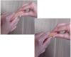
Metacarpophalangreal Joint SD ART
How would you perform this treatment?
Patient: Seated, standing, or spuine
Physician: Seated or Standing
- Physician evaluates the motion at the metacarpophalangeal joint in flexion, extension,
abduction, adduction, clockwise and counterclockwise
circumduction. - When a restriction is felt, gentle repetitive motion is
made through the restrictive barrier toward the anatomic barrier. - Continue articulation until somatic dysfunction is
alleviated. - Reassess

Proximal and Distal Interphalangeal Joint SD ART
How would you perform this treatment?
Patient: Seated, standing, or spuine
Physician: Seated or Standing
- Physician evaluates the motion at the proximal and distal interphalangeal joints in flexion, extension, abduction, adduction, clockwise and counterclockwise circumduction.
- When a restriction is felt, gentle repetitive motion is made through the restrictive barrier toward the anatomic barrier.
- Continue articulation until somatic dysfunction is
alleviated. - Reassess