Jacewicz/Foehring - Motor and Sensory CN's Flashcards

1
Q

32-y/o woman with left face droop, funny taste of food on the left, and pain in the pinna of the ear on the left. She also cannot wrinkle her forehead on the left, or open her eyelid against resistance.

Where is the lesion? What other symptoms might she experience if questioned by a knowledgeable observer? Most likely dx?

A
  • LESION: left CN VII
  • OTHER SYMPTOMS: hyperacusis (INC sensitivity to certain sounds), dry eye
  • DX: Bell’s palsy -> thought to most likely be a herpetic infection, with swelling of the nerve, but lyme disease and sarcoid can also do this
    1. These pts all partially recover bc peripheral N regenerates (recovery can be incomplete, or to the wrong targets)
2
Q

52-y/o man with cervical discectomy (sx correction of herniated disc) for radiating neck and arm pain on left. Anterolateral approach. Upon awakening, pt noted his voice was hoarse and reduced (muffled cough). No other neuro symptoms.

What is your anatomical dx?

A
  • Injury to left recurrent laryngeal N
  • NOTE: can also be injured by aortic aneurysm or mass in mediastinum; if reduced gag reflex or uvular deviation (to unaffected side), this is a sign the lesion is higher up (i.e., Vagus)
3
Q

19-y/o with trouble turning head to the right. NF-1. Symptoms began a few months ago, and were slowly progressive. Exam confirms complaints. No other neuro signs.

Where is the lesion?

A
  • Left spinal accessory N (turns head to opposite side)
  • REMEMBER: SCM innervated by ipsilateral CN XI
4
Q

37-y/o woman with dysarthria. Tongue protrudes to left when she sticks it out. Fasciculations on the left side too.

Where is the lesion?

A
  • Left hypoglossal N lesion
  • NOTE: this case had a metastasis of breast cancer that involved the hypoglossal canal
5
Q

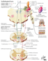
Name all of the cranial nerves that have a motor or PARA function, including their nuclei and function.

A
  • RED = voluntary
  • PURPLE = PARA
6
Q

Note the locations of the CN nuclei. You should know this shit by now.

A

Good job!

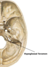
7
Q

Where is the motor nucleus of the trigeminal N? Where does it exit the calvarium, and what does it innervate?

A
  • Mixed motor and sensory N
  • Nuclei at MID-PONS level
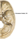
  • Motor fibers travel w/CN V via mandibular (V3) branch to exit calvarium via FORAMEN OVALE
  • Motor V innervates mm of MASTICATION, incl. the masseter and temporalis muscles
8
Q

Where does CN V exit the brainstem anatomically?

A
  • Mid-pons level of ventral surface of brainstem
9
Q

Where does Motor CN V exit the calvarium?

A
  • Motor CN V travels along floor of Meckel’s cave, and joins with V3 (mandibular) branch of trigeminal nerve
  • Exits the calvarium via the FORAMEN OVALE
10
Q

Describe the jaw jerk reflex.

A
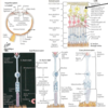
  • Tapping gently on lower jaw triggers muscle spindles in masseter muscle to send impulse via sensory fibers of sensory CN V, which synapse in MESENCEPHALIC nucleus of V
  • Short interneuron connects w/MOTOR nucleus of V to send an impulse to the masseter muscle to contract
11
Q

What do the motor fibers of the trigeminal nerve innervate?

A
  • Muscles of mastication: masseter and temporalis
  • Tensor tympani: dampens sound
  • Tensor veli palatini: opens eustachian tube
  • Mylohyoid: elevates hyoid bone
  • Anterior belly of the digastric: elevates hyoid
12
Q

How do you test motor CN V clinically?

A
  • Have pt bite down on tongue depressor and test jaw strength -> palpate masseter and temporalis mm
    1. Test bilaterally
  • Masseter muscle has mm spindles, and can be tested via muscle stretch reflex -> efferent arm of this reflex mediated by motor CN V
13
Q

How will UMN and LMN lesions to CN V manifest themselves?

A
  • UMN input to trigeminal motor nuclei largely bilateral, so unilateral lesions to motor cortex or corticobulbar fibers does NOT produce unilateral weakness of jaw opening or closing, but jaw jerk may be INC
  • LMN: lesions of CN V or its nuclei will cause unilateral weakness of jaw closure, reduced jaw jerk, and atrophy of temporalis and masseter mm on ipsilateral side
14
Q

Where are the motor/PARA nuclei of CN VII? Where do they exit the calvarium, and what do they innervate?

A
  • Mixed motor, PARA, and sensory N
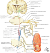
  • Motor component = facial nucleus (red box) at mid-pons level -> innervates mm of facial expression and several smaller mm, like the stapedius mm
  • PARA component = superior salivatory nucleus (purple box) near midline of rostral medulla -> sends preganglionic, PARA fibers through nervus intermedius to join CN VII to synapse in various ganglia with 2o innervation of lacrimal, salivary glands
15
Q

Where does CN VII exit the brainstem?

A
  • CN VII and nervus intermedius exit the brainstem at the pontomedullary junction in a region called the cerebellopontine angle
  • Ventral surface of the brainstem
16
Q

Describe the relationship of the facial and abducens nuclei/tracts.

A
  • Axons of CN VII initially pass dorsal-medially, and loop over abducens nucleus (red) before turning ventrally to exit brainstem at pontomedullary junction
  • NOTE: facial nucleus axons create a bulge on the floor of the fourth ventricle called the facial colliculus
17
Q

Where/how does CN VII exit the calvarium?

A
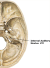
  • Facial N and nervus intermedius exit calvarium via auditory canal
  • Opening of this canal is called the internal auditory meatus -> w/in auditory canal, CN VII bends ventrally to enter the facial canal and exits the skull via the stylomastoid foramen
18
Q

Describe the anatomy of motor VII and nucleus intermedius, incl. their target organs.

A
  • Motor VII exits brainstem at pontomedullary junction, traverses cerebellopontine angle to enter internal auditory meatus and travel in auditory canal of petrous bone -> bends to enter facial canal, and exits cranium via the stylomastoid foramen
  • Fibers (nervous intermedius) of superior salivatory nucleus exit brainstem as small root adjacent to motor VII -> travel w/motor VII to synapse in sphenopalatine (pterygopalatine) and submandibular ganglia w/2o neurons innervating salivary/lacrimal glands
  • TARGETS: mm of facial expression, salivary/lacrimal glands
19
Q

What do the motor fibers of the facial N innervate?

A
  • MM of facial expression
  • Stapedius muscle: dampens sound
20
Q

How can you test motor CN VII?

A
  • Ask pt to wrinkle their forehead, close their eyes tightly, and show you their teeth
  • Look for symmetric furrowing of forehead, ability to symmetrically close eyes, and symmetric retraction of corners of the mouth
  • Motor VII and its nerve also mediate efferent arm of the corneal reflex -> gently stroke cornea, and observe eye closure
  • Also note width of palpebral fissure since weakness of orbicularis oculi m will cause widening of eye opening at rest (see attached image)
21
Q

How will UMN and LMN lesions to CN VII manifest themselves?

A
  • UMN input to motor CN VII largely bilateral for forehead mm, but unilateral for mm of lower face -> unilateral lesions to motor cortex or corticobulbar fibers causes unilateral weakness of contralateral lower face mm, but spares forehead mm (common cause is stroke)
  • Unilateral LMN lesions cause ipsilateral weakness of lower face AND forehead mm -> ipsilateral hyperacusis (sensitivity to certain sounds) and dry eye may also occur -> common cause is Bell’s palsy
    1. See attached image
22
Q

Where is the nucleus of CN IX? Where does it exit the calvarium, and what does it innervate?

A
  • Mixed motor, PARA, sensory N
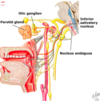
  • MOTOR: nucleus ambiguus (red box) near ponto-medullary junc; innervates stylopharyngeus m
  • PARA: inferior salivatory nucleus (purple box) near midline of medulla sends preganglionic, PARA fibers via IX to lesser petrosal N to synapse in otic ganglion w/2o innervation of parotid gland
23
Q

Where does CN IX exit the brainstem?

A
  • Exits at junction b/t pons and medulla
  • Ventral surface of brainstem
24
Q

Where does CN IX exit the calvarium?

A
  • Jugular foramen (w/X, XI, and internal jugular)
25
Q

Describe the anatomy of motor N IX, and its target organs.

A
  • Nucleus ambiguus fibers exit calvarium via jugular foramen and innervate stylopharyngeal m to raise pharynx during talking and swallowing
  • Efferent fibers from inferior salivatory nucleus travel w/CN IX, and split off as lesser petrosal N before synapsing in otic ganglion -> 2o neurons innervate parotid gland
  • TARGETS: stylopharyngeus m, and parotid gland
26
Q

How is CN IX function tested clinically?

A
  • Testing motor function difficult, and usually not done bc only innervates stylopharyngeal m
  • N integrity routinely tested under sensory func by testing gag reflex, of which CN IX mediates the sensory component (afferent):
    1. Motor component is shared by IX and X, but more so by X
    2. Tested by gently touching posterior pharynx on L and R separately, and watching for motor, or gag response, and elevation of soft palate
27
Q

Where is the nucleus of CN X? Where does it exit the calvarium, and what does it innervate?

A
  • Mixed motor, PARA, and sensory
  • MOTOR: served by nucleus ambiguus (red box) near lateral medulla, and innervates mm of soft palate, pharynx, and larynx
  • PARA: dorsal motor nucleus of Vagus (purple box) near midline of medulla sends preganglionic, PARA fibers to intramural ganglia associated with the heart, lung, and digestive tract (red arrow pts to nerve)
28
Q

Where does CN X exit the brainstem?

A
  • Series of rootlets between inferior olive and inferior cerebellar peduncle
  • Ventral surface of brainstem
29
Q

How does CN X exit the calvarium?

A
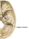
  • Jugular foramen (w/IX, XI, internal jugular)
30
Q

Describe the anatomy of motor nerve X.

A
  • Nucleus ambiguus fibers travel in CN X to innervate pharyngeal, laryngeal, and palate mm -> exits calvarium via jugular foramen
  • Preganglionic fibers of dorsal motor nucleus travel with CN X to synapse in intramural ganglia in walls of heart, lungs, and gut
31
Q

What do the motor fibers of Vagus innervate?

A
  • MM of the pharynx -> elevate pharynx during talking and swallowing
  • Also participates in efferent arm (motor component) of gag reflex (along with CN IX: sensory component)
32
Q

How will UMN and LMN lesions to CN X manifest themselves?

A
  • Unilateral UMN lesions tend to cause less prominent symptoms bc substantial bilateral UMN innervation of CN X nuclei
  • Unilateral lesions involving nucleus ambiguus or CN X will cause hoarseness, dysphagia, and inability to elevate palate on ipsilateral side
    1. Reduced gag reflex on stimulated side
33
Q

What do the PARA fibers of CN X innervate?

A
  • Dorsal motor nucleus of Vagus and PARA fibers influence many autonomic functions, incl. hear rate, respiration, and digestion
34
Q

Where is the nucleus of CN XI? Where does it exit the calvarium, and what does it innervate?

A
  • Pure motor N that controls:
    1. Head turning via innervation of SCM mm
    2. Shoulder elevation via trapezius mm
  • Receives motor axons from caudal portion of nucleus ambiguus (lateral medulla) & spinal accessory nucleus
    1. Spinal accessory nucleus made of neurons in intermediolateral gray area b/t dorsal and ventral horns of first 4-5 segments of cervical spinal cord
  • Fibers from spinal accessory nucleus exit cord b/t dorsal and ventral roots, and ascend alongside spinal cord -> enter calvarium via foramen magnum, and exit it via jugular foramen
  • NOTE: fibers originating from nucleus ambiguus travel short distance with CN XI before joining CN X to innervate larynx and pharynx (more appropriately considered part of X)
35
Q

Where does CN XI exit the brainstem?

A
  • CN XI axons exit lower medulla and upper spinal cord, joining together to form the spinal accessory N
36
Q

Where does CN XI enter and exit the calvarium?

A
  • Enters via foramen magnum
  • Exits via jugular foramen
37
Q

Describe the anatomy of motor N XI.

A
  • Caudal end of nucleus ambiguus sends fibers through proximal portion of CN XI before joining CN X
  • Fibers from spinal accessory neurons in C1-C5 cervical cord ascend through foramen magnum and exit via jugular foramen to innervate SCM (ipsilateral) and trapezius (contralateral) mm
38
Q

What do the motor fibers of CN XI innervate?

A
  • Innervate contralateral trapezius mm to elevate shoulder
  • Ipsilateral SCM to rotate head in the opposite direction
39
Q

How will UMN and LMN lesions to CN XI manifest themselves?

A
  • UMN: unilateral UMN lesions will cause weakness of contralateral shoulder elevation and weakness of ipsilateral SCM -> i.e., a left UMN lesion will produce weakness of right shoulder, and turning head to the right (left SCM)
  • LMN: unilateral lesions of either CN XI or its motor nuclei will produce weakness and atrophy of both mm ipsilateral to the lesion -> weakness of elevating ipsilateral shoulder and ipsilateral SCM turning head away from injured side
40
Q

Where is the nucleus of CN XII? Where does it exit the calvarium, and what does it innervate?

A
  • Pure motor nerve that controls tongue mvmt
  • Served by hypoglossal nucleus, which lies near midline in mid- to posterior portion of medulla
  • Axons exit brainstem ventrally b/t inferior olives and the pyramids
  • Note CN XII is not shown on this slide since it exits the brainstem on the ventral surface (see slide 34).
41
Q

Where does CN XII exit the brainstem?

A
  • Group of N fibers exiting brainstem b/t inferior olives and pyramids, then join to form hypoglossal N
  • Note location of N at junction of medulla and spinal cord
42
Q

Where does CN XII exit the calvarium?

A
  • Hypoglossal foramen: lies along posterior aspect of petrous temporal bone
43
Q

Describe the anatomy of CN XII.

A
  • Exits brainstem ventrally, and exits calvarium via hypoglossal canal to innervate tongue mm
44
Q

What does the hypoglossal N innervate? What happens in the case of a LMN lesion?

A
  • All intrinsic and extrinsic tongue mm
  • Tongue protrudes from mouth in midline for normal individuals -> contraction of tongue mm on left pushes tongue out mouth to the right, and vice versa
  • Unilateral lesions of CN XII or its nuclei (LMN) will produce tongue protrusion towards side of the lesion, unilateral atrophy, and fasciculations
45
Q

What happens in the case of UMN lesions to the hypoglossal N?

A
  • Corticobulbar fibers from motor cortex cross over to control contralateral CN XII nuclei -> UMN lesion will cause tongue to deviate away from lesion
46
Q

39-y/o woman presents to office with complaint of “excruciating, lance-like pain lasting seconds over right jaw for past 2 weeks.” Shooting pains can be set off by brushing teeth.

Where is the lesion?

A
  • Right mandibular branch of CN V, right spinal nucleus of V, or right N root
    1. Happens bc aberrant vessel embeds itself in brainstem at trigeminal entry point, compressing it w/each pulsation (branch of basilar, like AICA)
  • Can also get facial hemispasm -> different presentation
47
Q

45-y/o man with recurrent brief attacks of excruciating pain. Usually begins at back of tongue or throat, and sometimes spreads to left ear. He has fainted with two of these attacks. They can be brought on by chewing, coughing, swallowing, or sneezing.

Where would you place lesion?

A
  • L N root of IX -> glossopharyngeal neuralgia
48
Q

What is the early division in the devo of the spinal cord/neural tube?

A
  • Dorsal (posterior) alar plate: sensory (afferent)
  • Ventral (anterior) basal plate: motor (efferent)
  • Divided by sulcus limitans
49
Q

What is encompassed in this red box? How are the motor v. sensory nuclei arranged in the yellow cross-section?

A
  • RED = floor of the 4th ventricle -> sulcus limitans can be seen as outer margin of floor of 4th ventricle
  • In adult, neural tube has opened dorsally, and alar (sensory) plate is displaced laterally relative to basal (motor) plate
  • Cross-section of adult rostral medulla: dotted lines indicate sulcus limitans (border of floor of 4th ventricle), marking boundaries b/t alar (dorsal) plate and basal (ventral) plate
50
Q

What are the key sensory CN nuclei? Where are they located?

A
  • 3 sensory nuclei for the trigeminal nucleus:
    1. Mesencephalic: mesencephalon
    2. Chief sensory: pons
    3. Spinal: column from mesencephalon to spinal cord, where it is continuous w/substantia gelatinosa (layers I and II) -> several cranial nerves (V, VII, IX, X) project to this nucleus
  • PONS: vestibular and cochlear nuclei (concerned w/body position and hearing, respectively) of CN VIII
  • Several CN’s project to solitary nucleus (medulla) -> rostral part of nucleus concerned w/taste (CNs VII, IX, X) and the caudal part concerned w/cardiorespiratory inputs
51
Q

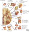
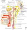
Briefly describe the locations and functions of the 12 CN’s (image).

A
52
Q

Briefly describe the anatomy and function of CN I. What happens if it is not functioning?

A
  • Completely sensory, and serves OLFACTION
  • Sensory receptors in nasal epithelium -> these cells send very fine axons (olfactory nerves or filae) through cribriform plate to synapse in olfactory bulb
  • Axons of projection neurons in bulb travel as olfactory tract to synapse in olfactory cortex, amygdala, and related forebrain structures
  • Anosmia: condition marked by loss of sense of smell
53
Q

What is the function of CN II? What is it made up of?

A
  • Optic N: completely sensory -> transmits visual info to the brain
  • Receptors are located in retina, at rear of eye -> optic N made of axons of retinal ganglion cells
54
Q

Describe the anatomy of the optic N -> what is its pathway/destination?

A
  • Optic N’s from e/eye converge just outside mesencephalon; at this pt, about 50% of fibers from e/eye cross to opposite side of body -> optic chiasm
  • Central to chiasm, fibers run as the optic tract, which runs along outside of mesencephalon to lateral geniculate body (the visual thalamus: LGN), which then projects to 1o visual cortex (banks of calcarine fissure)
  • Some optic N fibers bypass LGN and travel in the brachium (arm) of superior colliculus to synapse in the superior colliculus (tectum of mesencephalon) -> this input is important for visual startle reflexes
55
Q

Which branches of the trigeminal nerve supply what parts of the face?

A
  • V1: opthalmic
  • V2: maxillary
  • V3: mandibular
  • Carry fine touch, pain, and temp afferents from the skin of the face (note image for distribution)
  • NOTE: back of the head mostly innervated by spinal nerves from C2
    1. External ear (pinna) also innervated by multiple CN’s
56
Q

Describe the locations and functions of the trigeminal N nuclei.

A
  • CHIEF SENSORY: located in rostral pons, receives epicritic inputs from afferents for fine touch/vibration (analagous to posterior column nuclei)
    1. Cell bodies in trigeminal (Gasserian) ganglion outside brainstem (analagous to DRG) -> these cells synapse in chief sensory nucleus
    2. Fine touch afferents that run in o/CN’s (VII, IX, X) have cell bodies in different peripheral ganglia, but also synapse in chief sensory of V
    3. First integration center for all fine touch and vibration inputs for face
    4. Trigeminal lemniscus runs from chief sensory nucleus to VPM of thalamus (analagous to medial lemniscus for spinal pathways)
  • MESENCEPHALIC: cell bodies of proprioceptive afferents (mm spindles, GTO’s) from mm of mastication (and probs extraocular mm and mm of tongue)
    1. Essentially a displaced peripheral ganglion in tegmentum of mesencephalon -> peripheral processes of these cells form mesencephalic tract of V
  • SPINAL: extends from mesencephalon to spinal cord, where it is continuous w/substantia gelatinosa (layers I and II of dorsal horn)
    1. Receives inputs from pain/temp afferents for face that run in CN V (cell bodies in trigeminal ganglion), VII, IX, and X
    2. Central processes of cells in trigeminal ganglion project into and synapse in spinal nucleus of V; these axons and 2nd order axons from spinal nucleus of V run in spinal tract of V
    3. Trigeminothalamic tract runs from spinal nucleus of V to VPM (analogous to spinothalamic tract for spinal pathways)
57
Q

What are the associated tracts and functions of the 4 trigeminal nuclei (table)?

A
58
Q

Describe the somatotopic organization of the spinal tract and nucleus of V.

A
  • Afferents from the different CN’s and N branches stay segregated in spinal tract and nucleus of V (see figure with color coding)
  • V1, V2, V3 ventral to dorsal
  • Afferents from CN’s VII, IX, X most dorsal
  • NOTE: this pattern can be useful for localizing brainstem lesions
59
Q

Describe the trigeminal N sensory pathways.

A
  • Cell bodies of proprioceptive afferents from mm of mastication (+ extraocular mm and tongue) located in mesencephalic nucleus of V (GREEN) -> project bilaterally to trigeminal motor nucleus to mediate monosynaptic stretch reflexes
  • Cell bodies of afferents for fine touch and vibration are in ganglia outside brainstem (esp. trigeminal ganglion of V, but also geniculate of VII, inf/sup glossopharyngeal of IX, and inf/sup vagal of V) -> project to chief (principal) nucleus of V (RED), and 2nd order projections from this nucleus cross midline and travel in trigeminal meniscus to the VPM (thalamus)

The cell bodies of pain and temperature afferents (shown in blue) are located in various ganglia (Trigeminal for V, geniculate for VII, for inferior and superior glossopharyngeal for IX, inferior and superior vagal for X). Thus several cranial nerves contribute to this nucleus (V, VII, IX, X). Their central processes project into the spinal tract of V and synapse in the spinal nucleus of V. The second order projections cross the midline and travel in the trigeminothalamic tract (also shown in red).

60
Q

What are the 2 sensory components of CN VII? Where are the cell bodies, and where do they project to?

A
  • Taste sensation for anterior 2/3rds of tongue
  • Somesthetic afferents (fine touch, vibration, pain, temp) for small area around external auditory meatus
    1. Cell bodies in geniculate ganglion, and central processes project to chief sensory nucleus (epicritic) or spinal nucleus (pain/temp) of V
61
Q

What are the 4 nuclei, associated tracts, and functions of CN VII (table)?

A
62
Q

Where do taste fibers from the tongue project? Describe the relevant anatomy.

A
  • Taste fibers project to the rostral part of the nucleus solitarius (gustatory nucleus): column through pons and medulla
  • Gustatory nucleus receives input from VII, IX, X
63
Q

What is the function of the nucleus solitarius? Describe the relevant anatomy.

A
64
Q

What is the function of CN VIII?

A
  • The vestibulocochlear N has one functional component (sensory) with two divisions:
    1. Vestibular: balance and acceleration
    2. Auditory: hearing
65
Q

Where are the vestibular and auditory receptors located for CN VIII?

A
  • Sensory afferents for audition (hearing) & vestibular sense (body position in space) are in inner ear
  • AUDITORY receptors (hair cells) in organ of Corti in the cochlea -> sensitive to airborne pressure waves, as transduced into fluid waves in the cochlea
    1. Depending on location in cochlea, these receptors are tuned to different frequencies of sound (tonotopic org)
  • VESTIBULAR receptors are also hair cells, and are located in the saccule, utricle, & 3 semicircular canals
66
Q

Briefly describe the auditory pathways.

A
  • Cell bodies of auditory afferents are in the spiral ganglion, and send axons via CN VIII (cochlear division) to synapse in dorsal/ventral cochlear nuclei
  • Hair cells and supporting cells comprise organ of Corti in the cochlea -> synapse on spiral ganglion, and these cells send their axons in the cochlear division of the vestibulocochlear N
  • Cochlear N fibers synapse in ipsilateral dorsal and ventral cochlear nuclei (at pontomedullary junction)
  • Auditory pathways ascend bilaterally to inferior colliculus, and these neurons project to the medial geniculate body (auditory thalamus)
  • Medial geniculate projects to auditory cortex via Heschel’s gyrus, transverse gyri in superior temporal lobe
67
Q

Briefly describe the vestibular pathways.

A
  • Important for adjustment of posture, regulating mm tone, and coordination of eye mvmts
  • Hair cells in semicircular canals, saccule, and vestibule -> synapse in vestibular ganglion (Scarpa’s ganglion), and send their axons in vestibular division of CN VIII to synapse in vestibular nuclei (in pons and rostral medulla, near floor of 4th ventricle)
  • 4 vestibular nuclei:
    1. LATERAL: gives rise to lateral vestibulospinal tract, a descending motor pathway (balance, extensor tone)
    2. MEDIAL/INFERIOR: medial vestibulospinal tract extends to cervical levels of the cord, and helps control neck and head position
    3. MEDIAL/SUPERIOR: contribute to medial longitudinal fasciculus (MLF), which is important for coordinating eye movements
68
Q

What are the sensory components of CN IX?

A
  • Taste from posterior 1/3 of tongue
  • Somatic sensation from pharynx and posterior 1/3 of tongue
  • Chemo- and baroreception from carotid sinus and bodies
  • Fine touch, pain, temp from middle ear, and portion of external ear
69
Q

Which nuclei in the brainstem are associated with CN IX?

A
  • Afferents for taste from post 1/3 of tongue terminate in rostral solitary tract (gustatory nucleus)
  • Afferents from baro- (carotid sinus) and chemo-receptors (carotid body) project to caudal part of solitary nucleus and tract (cardioresp nucleus); sensory/afferent pathway of gag reflex also projects here
  • Pain/temp afferents from post 1/3 of tongue, pharynx, tonsils, tympanum, and small area around external ear project into spinal tract and nucleus of V
  • Afferents for epicritic touch from post 1/3 of tongue, pharynx, tonsils, tympanum, and small area around the external ear project to the chief sensory nucleus of V
  • NOTE: nucleus ambiguous is a motor nucleus containing neurons that project with CNs IX and X
70
Q

What is the nucleus that serves the CN IX baro- and chemoreceptors?

A
  • Baroreceptors (carotid sinus) and chemoreceptors (carotid body) project to caudal part of solitary nucleus and tract
71
Q

Describe the 7 nuclei (some repeat), and their associated tracts and functions for CN IX (table).

A
72
Q

What are the sensory components of CN X?

A
  • Taste from pharynx and epiglottis
  • Visceral info from aortic arch baro- and chemo-receptors
  • Somatic info from pharynx and larynx
  • Touch, pain, and temp from small portion of external ear
73
Q

What nuclei serve the sensory components of CN X?

A
  • Taste from pharynx and epiglottis afferents project to rostral solitary tract and nucleus
  • Afferents carrying visceral info from aortic arch baro- and chemoreceptors project caudal solitary tract and nucleus (NOTE: afferents w/general visceral sense from pharynx and larynx also project here)
  • Afferents carrying somatic info from pharynx, post meninges, and larynx, and touch, pain, and temp from small portion of external ear project to spinal tract and nucleus of V
74
Q

What are the 5 nuclei (some repeat), and their associated tracts and functions of CN X (table)?

A
75
Q

Describe the gag reflex pathways.

A
  • This reflex is for protecting and clearing airway in response to irritant to palate, pharynx, and associated areas
  • Afferent limb carried by afferents of CN IX projecting to caudal solitary nucleus
  • Interneuron is interposed b/t afferent and efferent in nucleus ambiguous -> efferent response is mediated by motoneurons in nucleus ambiguus, whose axons travel with CN X