Jacewicz - Cerebellum and Brainstem Flashcards
(33 cards)
Describe general cerebellar function.
- Comparator that compensates for error in mvmt by comparing intention with performance -> anticipates and smooths out mvmts of trunk and limbs
- Coordination of somatic motor activity, regulation of mm tone, & mechs that influence/maintain equilibrium
- Contributes to non-motor func, like: cognition, emotion, affective processing
- Plays a role in sequencing incoming sensory patterns and detecting temporal changes in sequence of sensory events
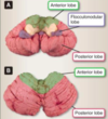
Describe the surface anatomy and homuncular distributions of the cerebellum.
- Vermis = pink; paravermian area = orange
1. Control axial musculature (neck and trunk mm) - Cerebellar hemispheres = green (REMEMBER: there is an anterior, and a posterior lobe)
1. Control the limbs (arms and legs) - Flocculonodular lobe = blue
1. Heavily involved in maintaining balance and in coordinating head/eye mvmts w/vestibular system

Where are the deep cerebellar nuclei located anatomically (image), and what areas of the cerebellum project to them?
What happens if they are damaged?
- These nuclei serve as 1o relay pts for efferent fibers from cerebellar cortex (Purkinje cells) to o/brain regions
- Lateral hemispheres project to DENTATE nuclei
- Paravermal zones project to GLOBOSE/EMBOLIFORM nuclei, collectively known as the interpositus nuclei
- Vermis projects to FASTIGIAL nuclei
- NOTE: damage to these deep cerebellar nuclei cause severe ataxia, far worse than the ataxia arising from damage to the much larger cerebellar hemispheres

What are the 3 fiber bundles that carry afferent/efferent nerve fibers to/from the cerebellum? Name the afferent and efferent inputs for all 3.
- SUPERIOR cerebellar peduncle:
1. Afferents: anterior spinocerebellar tract, acoustic and optic information
2. Efferents: dentatorubrothalamic tract, dentatothalamic tract - MIDDLE cerebellar peduncle:
1. Afferents: pontocerebellar tract - INFERIOR cerebellar peduncle:
1. Afferents: vestibulocerebellar tract, olivocerebellar tract, post spinocerebellar tract
2. Efferents: cerebellovestibular tract, cerebelloolivary tract

How is cortical motor “intent” relayed through the cerebellum?
- Cortical motor intent sent to nuclei in pons that forward the info to contralateral cerebellar hemisphere for processing -> enters via MCP
- Once processed, info sent back to cortex via dentate nuclei -> SCP is major outflow pathway to forebrain via dentatorubrothalamic and dentatothalamic tracts
- Dentatothalamic fibers carry info from lateral portions of ant and post cerebellar cortices to contralateral VL of thalamus -> then to motor cortex to smooth out mvmt in limbs ipsilateral to cerebellar hemisphere of origin
- Final common pathway for this coordinated movement is through the corticospinal tract
What pathways flow through the ICP?
- Carries proprioceptive input from post spinocerebellar tract, cerebellovestibular, and cerebelloolivary fibers
- CV and CO carry info from vermis and flocculonodular lobes through emboliform, globose, and fastigial (EGF) nuclei to vestibular nuclei, olivary nuclei, and brainstem reticular formation
- These pathways are important in maintaining balance
- Note the attached image of the cerebellar surface anatomy

What are these three layers? Name the 5 different neuron types in the cerebellar cortex/grey matter.

- LAYERS: molecular, purkinje, and granular
- NEURON TYPES: Basket and Stellate cells (outer layer), Purkinje cells (middle layer), Golgi and Granule cells (granule layer)

What are the Purkinje cell connections?
- Only direct input (afferent) to Purkinje cells from outside are CLIMBING FIBERS (origin in contralateral olivary nuclei; tremendous influence on Purkinje cell)
- Other input from outside the cerebellum via MOSSY FIBERS that first synapse in cerebellar glomeruli
1. Synapse w/Granule and Golgi cell dendrites, and Golgi axon terminals
2. Granule cells pass modified info to Purkinje cell - Stellate/Basket cells have INH effect on Purkinje cells
- NOTE: Purkinje cells are the only output neurons of the cerebellar cortex; synapse on one of deep nuclei that send efferent fibers outside the cerebellum
1. Purkinje cells also have a large soma, so they more sensitive to hypoxia/ischemia

What are the 3 functional divisions of the cerebellum? What do they include?
- VESTIBULOCEREBELLUM: vestibular nuclei, flocculonodular lobe, inferior portion of paravermis, fastigial nuclei (oldest part; archicerebellum)
- SPINOCEREBELLUM: anterior lobe, vermis, superior paravermis (paleocerebellum; next oldest part)
- CEREBROCEREBELLUM: lateral portions of posterior lobes (neocerebellum)
What are the afferents, efferents, and function of the vestibulocerebellar system? What happens if this system is destroyed?
- AFFERENTS: from ipsilateral vestibular nuclei (in brainstem) via ICP -> project to flocculonodular lobe and inferior paravermal area
- EFFERENTS: to vestibular nuclei via fastigial nuclei and ICP -> info sent down spinal cord in vestibulospinal tract to exert truncal stability and balance
- FUNCTION: coordinate eye, head, neck mvmts, and maintain body balance -> destruction of this largely midline cerebellar system by stroke or disease causes severe truncal and gait ataxia
- NOTE: both feed-forward and feedback loops through cerebellum from both motor and vestibular systems, providing continuous correction to and anticipation of changes in body’s axial stability and balance

What are the afferent inputs to the spinocerebellum?
- PERIPHERAL LIMB COORDINATION: T1a and T2 fibers from mm spindles + T1b fibers from GTO’s carry proprioceptive info to dorsal horn of spinal cord -> synapse on Clarke’s column (lower limb) and accessory cuneate nucleus (upper limb), and take info via spino-cerebellar tract to ipsilateral anterior cerebellum via ICP
- MAINTAINING POSTURE of LOWER LIMBS: spinal border neurons (near border of lateral ventral horn of lower thoracic and lumbar spinal cord) receive input from higher centers like LMN’s, and send “copy” of motor instructions back to anterior cerebellum
1. Send axons via anterior (ventral) spinocerebellar tract, which access ant cerebellum via SCP

What are the efferent outputs of the spinocerebellum? What happens in the case of a lesion to this area?
- TRUNCAL: rubrospinal, vestibulospinal, reticulospinal
1. Processed primarily in vermis, and output via fastigial nucleus -> bilateral projections sent to vestibular and red nuclei, and reticular formation, then to spine via tracts listed above - LIMBS: EGF, VL (thalamus), motor cortex, corticospinal
1. Coordinated in anterior lobe, and output via emboliform and globose nuclei to VL of thalamus, which projects to motor cortex - LESION to ant cerebellum by stroke typically leads to truncal instability + peripheral limb incoordination

How is limb coordination lateralized in the cerebellum?
- Ipsilateral control
- Inferior olive provides info to cerebellum on mvmt from contralateral side, allowing synergistic limb mvmts while maintaining stability of the trunk
What are the functional details for the cerebrocerebellum? Function?
- Lateral aspects of posterior lobes; receive input from many areas of the cortex via pontine nuclei -> send fibers to contralateral cerebellum via MCP
- Output from CC primarily to dentate nucleus, which projects to red nucleus, then to VL of thalamus via dentatorubrothalamic tract (also a parallel direct path to thalamus from dentate via dentatothalamic tract)
- Important for eye-hand coordination needed to reach or manipulate an object: compares past sensory experiences to learn/predict sensory consequences of current mvmts (why you can’t tickle yourself)
- Important in planning and making voluntary mvmts automatic, like handwriting, typing, or piano playing
1. Also automatizes aspects of cognition: fluidity of language, automatic syntax and grammar, and prediction of sentence structure and flow

What deficit does this illustrate?

- Effect of prism glasses on normal (left) vs. cerebellar (right) pt
- Ability to learn to alter dart-throwing technique requires correct analysis of visual sensory info and motor output:
1. Normal person adjusts throws to become more accurate after a little practice
2. No compensation by pt with cerebellar degenerative disease
What are the clinical signs of cerebellar dysfunction?
- Unstable gait/stance w/tendency to fall; broad-based gait (sailor’s gate, reeling and drunken)
- Mvmts jerky, unsmooth + intentional tremor
- ATAXIA (dis-coordination) of trunk and/or extremities
- Dysmetria of mvmt: goal-directed mvmt can over- or undershoot target
- Eye mvmt disorders: nystagmus, saccadic and smooth pursuit dysmetria
- Speech disorders: ataxic dysarthria w/scanning speech, difficulty maintaining speech rhythm, intonation, and correct articulation
Identify the structures labeled in yellow, and their functions. What level is this?

- MIDBRAIN
- SUPERIOR COLLICULUS: control of reflex mvmts that orient eyes, head, neck in response to visual, auditory, somatic stimuli -> eyes automatically note peripheral ad that jiggles on website while you ignore ads that don’t
- PAG: enkephalin-producing cells that suppress pain; axons from reticular activating system pass through this area, with coma resulting if damaged
- CEREBRAL AQUEDUCT of SYLVIUS: passageway connecting third and fourth ventricles; some ppl born w/smaller caliber aqueduct that INC likelihood of hydrocephalus due to blocked CSF flow if meningitis
- EDINGER-WESTPHAL NUCLEUS: PARA innervation to constrict pupil, and to ciliary mm to alter lens shape for accommodation
- OCULOMOTOR NUCLEUS/NERVE: eye mm motor control
- SPINOTHALAMIC TRACT: ascends to thalamus w/pain and temp info from periphery
- MEDIAL LEMNISCUS: vibration and proprioception info to thalamus from contralateral nucleus gracilis and cuneatus where axons in posterior columns terminated
- MEDIAL GENICULATE BODY: thalamic relay nucleus for auditory information ascending along multiple way stations in the brainstem
- LATERAL GENICULATE BODY: thalamic relay nucleus for visual information (receives 85% of optic tract fibers, while about 15% terminate in superior colliculus nuclei)
- CEREBRAL PEDUNCLE: fiber bundles of corticospinal tract connecting cerebral cortex to brainstem (face, arm fibers medial, leg fibers lateral, like internal capsule)
- OPTIC TRACT: emerges from the optic chiasm and 85% of its fibers end in the lateral geniculate body
- SUBSTANTIA NIGRA: closely involved w/basal ganglia that modulates motor mvmt
- RED NUCLEUS: relay station for cerebellar projections to thalamus (pale pink in autopsy specimens due to ferritin and hemoglobin)
- MEDIAL LONGITUDINAL FASCICULUS (MLF): midline fiber pathway connecting vestibular nuclei and CN nuclei III, IV, VI to coordinate head and eye movements
Identify the structures labeled in yellow, and their functions. What level is this?

- PONS
- SUPERIOR CEREBELLAR PEDUNCLE: chief pathway for cerebellum to send processed info to thalamus (VL) and motor cortex for implementation (ant cerebello-spinal tract enters cerebellum here)
- MIDDLE CEREBELLAR PEDUNCLE: receives fibers from contralateral pontine nuclei, where fibers from cortical motor centers terminate to access cerebellum
- MESENCEPHALIC, MAIN SENSORY, MOTOR NUCLEI of CN V: note that mesencephalic nucleus originates in mesencephalon/midbrain, but extends into pons
- PONTINE NUCLEI: collection of neurons in pons that receive input from neocortex and send crossing fibers via MCP
- LOCUS COERULEUS: noradrenergic brainstem nucleus involved in emotion, arousal, sleep/wake cycle
- RAPHE NUCLEI: mood, vigilance, and levels of alertness, sleep/wake cycles -> release serotonin to rest of the brain (SSRI’s thought to act on these nuclei and their targets)
- CORTICOSPINAL TRACT: carries motor fibers from neocortex to spinal interneurons and LMN’s
Identify the structures labeled in yellow, and their functions. Where is this?

- UPPER MEDULLA
- INF/MEDIAL VESTIBULAR NUCLEI: regulate balance
- NUCLEUS/TRACTUS SOLITARIUS (NTS): sensory nuclei for taste (CN VII, IX, X) and various chemo- and baroreceptors (CN IX, X) -> important for gag, carotid sinus, aortic, cough, baro/chemoreceptor, respiratory reflexes and regulation of GI secretion/motility
- DORSAL MOTOR NUCLEUS OF VAGUS (CN X): PARA motor nucleus to lungs and gut
- DESCENDING SPINAL NUCLEUS/TRACT of CN V: receives info about crude touch, pain, temp from ipsilateral face via input from CN V, VII, IX, X, and extends down upper cervical spinal cord, where it merges w/substantia gelatinosa of dorsal horn
- INFERIOR OLIVARY NUCLEUS: sends climbing fibers that synapse on cerebellar Purkinje cells
- PYRAMID: corticospinal tract fibers in medulla
- RETICULAR FORMATION: network of neurons and axons that reside in brainstem tegmentum, and are involved in arousal (midbrain and upper pons), respiration, and HR control in medulla
Identify the structures labeled in yellow, and their functions. Where is this?

- LOWER MEDULLA
- FASCICULUS CUNEATUS/GRACILUS: proprioception and vibration info from arms/legs, respectively
- DECUSSATION: descending corticospinal tracts cross here; arm fibers decussate first (medial portion of upper cervical corticospinal tract), and leg fibers decussate more caudally (lateral portion of upper tract)
1. Lesion at the decussation can produce “crossed paralysis,” like left leg, right arm by involving uncrossed arm fibers and crossed leg fibers on one side (right, in this example) -> rare, and more often, quadriparesis followed by death result from a severe lesion at this level
Identify the nerves/nuclei. What do the colors indicate?

- Sensory CN nuclei in blue
- Motor CN nuclei in red
- PARA nuclei in purple (right)

What is a common thread to most of the brainstem syndromes? What might cause them?
- COMMON THREAD: pattern of contralateral body weakness or sensory loss, coupled with ipsilateral cranial nerve weakness or sensory loss
- MCC focal vascular occlusions and stroke, but other brain disorders may also cause these syndromes
- NOTE: territory supplied by individual aa and their branches varies considerably from person to person, so it is common for these syndromes to be smaller and incomplete, or larger and include deficits from neighboring areas
What will the syndromic affects of this lesion be? Describe brainstem region, arterial supply, structures involved, and the clinical signs/symptoms.

- BRAINSTEM REGION: base of midbrain
- ARTERIAL SUPPLY: tip of basilar artery, and/or branches of PCA
- STRUCTURES INVOLVED: CN III fascicles, cerebral peduncle (corticospinal/corticobulbar tracts)
- CLINICAL SIGNS/SXS: ipsilateral CN III paresis with wide fixed pupil, contralateral hemiparesis, and contralateral facial and tongue weakness (lower face weaker than upper; tongue protrudes to weaker side)

What will be the syndromic affects of a lesion to the midbrain tegmentum? Describe arterial supply, structures involved, and the clinical signs/symptoms.
- ARTERIAL SUPPLY: tip of basilar artery, and/or branches of PCA
- STRUCTURES INVOLVED: CN III fascicles, red nucleus, superior cerebellar peduncle (dentatothalamic tract) +/- medial lemniscus, substantia nigra (exact location and extent of lesion unresolved)
- CLINICAL SIGNS/SXS: ipsilateral CN III paresis, contralateral tremor and ataxia


