heme/onc Flashcards

1
Q

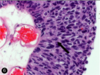
pt presents w -weight loss, night sweats, cough -CXR= apical granulomas w areas of central necrosis surrounded by large cells with abundant pale cyptoplasm most likely dx what are the large cells w pale cytoplasm? what cell marker are they most associated w

A

TB (apical granulomas = central caseous necrosis surrounded by Møs) macrophages = associated w CD14

2
Q

pt w sickle cell comes in for fever, dyspnea, chest pain: prior hospitalizations for abd pain were trx w hydration and resolved low hct, high reticulocyte they die in the hospital spleen is found to be firm and brown what is the dx? explain the spleen findings

A

sx= Acute Chest Syndrome : a vaso-occlusive crises localized to the pulmonary vasculature that can occur in sickle cell patients = commonly precipitated by a previous lung infection -occurs in pts w homozygous HbS = repeated events of vaso-occlusion produce a spleen that is shrunken and fibrotic -spleen is brown bc of hemosiderosis secondary to years of extravascular hemolysis -this is the reason that most homozygous sickle cell patients have undergone “autosplenectomy” by the time they reach adulthood

3
Q

tissue factor aka ____ plays a significant role in what ds

A

tissue factor aka thromboplastic associated w DIC: will cause excessive production of thrombin + widespread intravascular fibrin deposition and fibrinolysis –> consumption of clotting factors and platelets

4
Q

most head and neck carcinomas will met/spread via what method

A

LYMPH (not direct extension) i.e. tonsils –> jugular LNs

5
Q

chemo therapy w a cyclophosphomide (a _____-based chemo) has an increased risk of what AE in F patients how will it present how can it be prevented

A

chemo trx w a nitrogen-mustard based chemo i..e a cyclophosphamide –> HEMORRHAGIC CYSTITIS : inc frequency, dysuria, suprapubic pain, progressive hematuria –> prevent this w aggressive hydration and cotrx w MESNA (2-mercaptoethanesulfonate) that will bind the toxic metabolites of the drug in the urine

6
Q

in sickle cell anemia, the hemoglobin of cells aggregates in response to what

A

“sickling” = aggregation of hemoglobin molecules in response to anoxic conditions

  • after polymerization, HbS forms a meshwork of fibrous polymers –> RBC distorts into the abn sickle shape

sickling is promoted by…

  • low O2 levels
  • inc acidity
  • low blood volume (dehydration)

(aka the conditions seen in the periphery –> inc sickling in the periphery –> impede BF–> microinfarcts all over the body–> vasoocclusive crises)

-organs that are predisposed to more sickling are

  • organs where blood moves more slowly –> low O2 and acidic conditions
    • spleen and liver
  • organs w high metabolic demand = inc O2 unloading
    • brain, M, placenta
7
Q

compare and contrast the action of unfractioned heparin and LMWH on

  • factor Xa
  • thrombin

give an example of a LMWH

A

thrombin = factor I

LMWH= enoxaparin

8
Q

what is porphyria cutanea tarda

  • etiology
  • presentation
    *
A

is the MC disorder of porphyrin (heme) synthesis

(porphyria= deficiency in teh early steps of heme synthesis that result in inc of porphyrin-containing metabolites)

  • etiology: x uropophyrinogen decarboxylase
    • any loss of enzyme after PBG conversion (aka late step dysunction) [see picture] wll result in PHOTOSENSITIVITY bc of the build up of UV LIGHT SENSITIVE PORPHORINS
  • presentation:
    • acquired > inherited
    • risk factors: inc alc intake, smoking, hep C, HIV
    • photosensitivity= blisters and vesicle formation on sunexposed areas (i.e. dorsum of hand) : small itchy spots that will blister–> rupture–> heal with hyperpigmentation
    • +edema, pruritis, erythema, pain
9
Q

what is the difference in a heme synthesis defieciency and a porphyria

A

porphyria: an enzyme deficiency at specific points in the heme synthesis pathway that result in the build up of porphyrins that are UV light sensitive

  • late step deficiencies= any enzyme after PBG (PBG deaminase, UP III synthase, UP decarboxylase, CP oxidase
  • =photosensitivity= blistering in sunexposed areas + pruritis, hyperpigmentation, erythemia, and pain

heme synthesis deficiency is an xferrochetalase or ALA synthase

  • this just presents as a microcytic, hypochromic anemia
10
Q

child presents w HA and gait ataxia

what is the most likely brain tumor

  • classic presentation?

differentiate this from the other common brain tumor in children

A

pilocytic astrocytomas are the most common brain tumor in children

  • HA and cerebellar sx
  • imaging shows well demarcated solid and cystic components
  • H&E = GFAP+ w hairlike processes and classic rosenthal fibers

VS MEDULLOBLASTOMA

  • =the most common malignant tumor in children
  • they are exclusively in the cerebellum
  • NOT cystic like pilocytic astrocytomas
  • imaging will show a solid mass in the cerebellum
  • the mass can compress on the v4th ventricle and lead to hydrocephalus (on imaging will look like a huge fluid build up in the superior ventricular system)
  • H&E: small cells with hypochromatic nuclei that form homer-wright rosettes
11
Q
  • which drug class will inc PT+PTT but have NO effect on thrombin time
  • which drug class will have an effect on thrombin time
  • which pathway must be inhibited to effect PT time
  • which pathway must be inhbited to effect aPTT time
A
  • direct Xa inhibitors
  • direct thrombin inhibitors
  • extrinsic
  • intrinsic
12
Q

what are CD3+ cells and what role do they play in the stem cell transplant process

A

CD3+ = CD4/CD8+ T cells

they are the cells in the donor BM that can cause graft-vs-host ds

(are often depleted from the graft before transplant)

13
Q

what is the MOA of hydroxyurea

what is its use in sickle cell ds

A
  • hydroxyurea
    • has myelosuppressive effects by arresting cell cycle in rapidly dividing hematopoetic cells
    • will inc the HbF levels
  • effects
    • improve O2 delivery
    • reduce vasoocclusive crises
    • lessen need for transfusions
14
Q

acute intermittant porphyria

  • etiology
  • presentation
  • trx
A
  • =PBG deaminase deficiency
  • hx of abd pain&gen GI sx + neuro sx (change in sleep/concentration, anxiety, numbness+tingling)
    • +change in urine (port wine color)
  • trx= ALA synthase inhibition
    • long term management w glucose or heme will also inhibit ALA
15
Q

how does HPV associated CA present

what gene mutation does HPV cause

A
  • HPV related CA– sore throat, enlarged/ulcerated tonsil, with a neck mass (often firm and immobile bc malignant)
  • HPV is associated w HEAD AND NECK CA
  • HPV release E6 and E7 proteins
    • E6 protein of HPV degrades p53 from the TP53 gene
    • E7== x Rb protein tumor suppressor) from the RB gene
16
Q

what is the similarity between RET and BRAF

what tumors are they associated with

A

both are protooncogenes that require GOF mutations to become oncogenes

BRAF

  • code fore Ser/Thr specific protein kinase
  • melanoma, NHL, papillary thyroid CA

RET

  • code for an RTK
  • MEN 2A and MEN 2B, papillary thyroid CA
17
Q

inc 5-HIAA in the urine represents what

A

the presence of carcinoid tumor

18
Q

why does warfarin need a bridge therapy w heparin till the INR levels out

having a heparin bridge lowers the risk of what AE

A

bc warfarin inhibits carboxylaton of NEW k-dependent clotting factors, your body first needs to use up all the pre-existing clotting factors in the plasma

only once those are all gone will the INR reach therapeutic activity

  • a bridge will lower the risk of skin necrosis due to the loss of Protein C before anything else (short half life)
19
Q

compare the change in PT, PTT, platelets, and bleeding time in…

  • hemophilia
  • vWF deficiency
  • DIC
  • uremic platelet dysfunction
  • heparin use
  • warfarin use
  • immune thrombocytopenia
A
20
Q

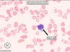
what are the pathologic cells seen in this histo pic

what is the dx?

clinical presentation

A

spherocytes

  • 2/3 the diameter of a normal RBC
  • more densely hemoglobinized around the periphery
  • no central zone of pallor
  • hereditary spherocytosis = x ankyrin and spectrin –> inc sequestration and destruction in the spleen
  • inc extravascular hemolysis = jaundice, splenomegaly, inc INDIRECT bilirubin
    • often young age
    • infection can lead to an acute hemolytic crisis –> acute worsening of pallow, scleral icterus, fatigue, and splenomegaly
21
Q

describe what an osteoblastic vs osteolytic bone lesion would look like

which metastatic CAs to the bone result in which

A

osteoblastic = SCLEROTIC = usually more indolent

osteolytic = LUCENT = tend to be from more aggressive CAs

22
Q

what are the bone changes seen w age

BE SPECIFIC

A

inc quantity of fat

dec overall mass

23
Q

desscribe the cause of abn bleeding in patients with uremia

how is this treated

A

pts w uremia (often ESRD, need dialyses) have abn platelets (qualitatively)

this results in n PT/PTT/platelet count but an abn bleeding time bc platelets are non-functional

treat w dialysis to remove the uremic toxins and allow the partial reversal of the abn

24
Q

what are the 5 features of anaplastic tumors

A
  • poorly differentiated
  • sign variation in size and shape of cells
  • abnormally large, hyperchromiatic nuclei
  • lots of mitotic figures
  • giant, multinucleated tumor cells
25
rapidly growing tumor of the mandible/jaw with palpable regional lymphadenopathy often an immigrant from the middle east - dx - histo findings - genetic mutations and associated causes - prognosis
- burkitt lymphoma - lymphocytes a basophilic cytoplasm, high mitotic index, diffuse distribution of **tingible body macrophages** (macrophages that phagocytose the cell debris) surrounded by clear space --\> **_starry sky appearance_** - assocated a c-Myc oncogene overexpression - v aggressive, but respond well to chemo
26
differentiate the mechanism of anemia causing disease in an M. pneumonia infection vs SLE by what mechanism does treatment relieve the anemia?
M. pnuemonia * cold agglutin * M. pnuemonia attacks respiratory epithelium with the I Ag --\> the body create IgM Abs, that also happen to be cross reactive in cold Temps--\> cause agglutination of RBCs--\> set off complement--\> intravascular hemolysis * trx--\> fall in IgM Ab creation will stop the cascade leading to hemolysis SLE * warm agglutination * IgG Ab bind to RBC membranes, causing them to be extravascularly hemolysed in the spleen * immune suppression will dec the IgG Ab creation
27
drugs that alter warfarin therapeutic levels (INR) in the body can do so by 3 mechanisms: (which drugs are in each category and what is the effect on INR/warfarin levels) * disruption to gut flora * activate CYP450 * inactivate CY450
* disruption to gut flora * **G (-)** gut flora make Vit K during their metabolism so abx that reduce Vit K producing gut flora will --\> **supratherapeutic INR (too much warfarin activity)** * abx that target G- bac= metronidazole, macrolides (azithryo, clarythro, erythro-mycin), flouriquinolones (floxacins) * activate CYP450 * --\> make the CYP450 metabolize warfarin faster --\> **subtherapeutic warfarin levels** * phenytoin, carbamezapine, rifampin * inactivate CY450 * --\> slow down the metabolism of warfarin--\> **supratherapeutic INR/warfarin levels** * metronidazole, fluconazole, amiodarone
28
this person has hemolytic anemia what three types of erythrocytes are seen in this pic and what are the sign of each
* polychromatic RBCs * =reticulocytes that are bigger than normal RBCs, slighly bluish, and lack a central pallor * indicate BM response to anemia * spherocytes * erythrocytes w reduced cell membrane * associated w immune mediated anemia * nucleated erythrocytes * are v immature RBCs * indicates a strong BM response to anemia *
29
what is the effective trx for an overdose in warfarin vs heparin
warfarin * immediate= FFP * over time: Vit K replenish heparin * protamine sulfate= a direct antagonist
30
what is primary myelofibrosis * etiology * presentation + findings
**primary myelofibrosis** = a _hematopoeitic stem cell malignancy_ in the BM that is associated w the _expansion of megakaryocytes_ * neoplastic megakaryocytes will secrete TGF-beta--\> stimulate BM fibroblasts to fill the medullary space w collagen --\> **bone marrow fibrosis** * this results in (**exramedullary hematopoeisis**) and **dacrocytes** on peripheral blood smear * present * _splenomegaiy and hepatomegaly_ * _anemia / pancytopenia_ secondary to EMH not being as efficienct as hematopoeisis in the BM * _tear drop cells and nucleated RBCs_ will appear in periphery as RBCs have to squeeze out of fibrotic BM/congested spleens * **dry tap on BM**: no marrow is yielded on BM aspiration due to all the fibrosis * old person, fatigue, unintensional weight loss, etc
31
what is the MOST important prognostic factor in urothelial (transitional cell) carcinoma
TUMOR DEPTH OF INVASION (even more than atypia or grade of the lesion i.e. an in situ lesion can have high grade) show in picture is an atypical mitotic figures (not the most important factor in prognosis, but def a prognostic factor)
32
alcoholism that develops over the course of WEEKS can cause an anemia by depleting what vitamin * presentation * etiology what other big Vit can be depleted by alcoholism and this cause anemia * how long would that take * what are other causes of deficiency * presentation
FOLATE: def develop over weeks * etiology: x nucleoside synthesis --\>x DNA production in blood cell precursors--\> abn cell division --\> megaloblastic hyperplasia of the BM (v big RBCs) * \*\*\*normally it would take months to become folate deficienct bc of body stores but an ALCOHOLIC can become folate deficient in weeks\*\*\* * peripheral blood smear shows **pancytopenia** and **hypersegmented neutrophils** (picture attached) * RBC abnormalities= ovalocytosis (elipses shape) and macrocytosis (iMCV\>100) vs Vit B12 (cobalamin) deficiency * this would take YEARS, even in an alcoholic * associated w pernicious anemia, chronic gastritis, certain meds, and fish tapeworm * presents w neuro sx (subacute combined degeneration w parasthesias ataxia, loss of proprioception)
33
differentiate between the two types of heparin induced thrombocytopenia (HIT)
* HIT Type 1 * a NON-IMMUNE mediated condition caused by platelet clumping * presents aruond 2 days after starting therapy * thrombocytopenia is only minimal (nadir of about 100k) * does NOT cause thrombosis (i.e. DVT/PE), so essentially nbd and can continue heparin * HIT Type 2 * Ab aggregation on the RBCs secondary to IgG Abs created against an heparin induced neoangtigen (**heparin platelet factor 4**) * manifests around 5-10 days after starting therapy * splenic Mø removal of Ab-marked RBCs results in significant thrombcytopenia * Ab-aggregation also results in widespread platelet aggregation--\> thrombocytopenia worsens --\> inc risk of thrombosis (venous or arterial) * pt may present with DVT/PE and thrombocytopenia shortly after beginning heparin therapy * NEED TO STOP HEPARIN IMMEDIATELY and switch to a different anticoag (i.e. argatroban)
34
what is a biologic response modifier? what are the MOA of the following BRMs and when are they used? * Rituximab * trastuzumab (aka herceptin) * infliximab * interleukin 2 * imatinib * abciximab
BRMs= treatments that are meant to boost the host immune response to better fight off a ds * Rituximab * a mab that targets the CD20 surface Ig * used in lymphoma immunotherapy * trastuzumab (aka herceptin) * anti-HER2-R mab * breast CA * infliximab * in IgG1 Ab against TNF-alpha * trx RA, ankylosing spondylitis, and fistulizing Crohns ds * interleukin 2 * a cytokine that regulates the differentiation of T cells to aid in tumor destruction * RCC and melanoma * imatinib * a potent inhibitor of the BCR/ABL protein tyrosine kinase found on the **philidelphia chromosome**, without causing cell apoptosis * trx CML * abciximab * mab against platelet GpIIb/IIIa receptor: blocks the final step in platelt aggregation * often administered during angioplasty in patients w ACS
35
multiple myeloma should be suspected with what patient presentation * what is the etiology of renal ds in pts with MM and how will be detected
suspect MM in an elderly patient with * some combination of **osteolytic lesions, anemia, hypercalcemia, or acute kidney injury** * AKI is caused by the deposition of lambda light chains of Igs in the PT of the kidney at levels that oversaturate the kidney's ability to reabsorb them * this results in them to combine, form obstructive casts --\> tubular rupture and kidney injury * monoclonal Abs will not be found on dipstick (bc they're not albumin) but they will show elevated protein on 24 hour urine
36
what is the genotypic change seen in sickle cell ds, (AA change and the codon change)
glutamic acid --\> valine GAG--\> GTG
37
explain how each of the results of sickle cell ds will present * hemolysis (labs) * vasoocclusive sx * children vs adults * infections
* hemolysis (labs) * =intra and extravascular --\> ↑INDIRECT bili, ↑ LDH, ↓ haptoglobin * (on change in the complement factor levels) * vasoocclusive sx * pain w hypoxic injury and ischemia: typically of BM, periosteum, and deep Ms * in **children**, oft present as **hand-foot syndrome**: dactylitis 2° infarctions of the bones in the extremities--\> swelling, edema, tenderness of the hands, feet * in **adults**: the small bones no longer contain hematopoeitic cells so they present with **acute chest syndrome, pain crises, leg ulceration, priapism, autosplenectomy, and stroke** * infections * w encapsulated organisms secondary to autosplenectomy * i..e S. pneumoniae
38
young boy with hx of bone CA presents w lung mass with the following histology dx?
ewing sarcoma * common in young children * mets to the lung and other bones EARLY ON * histo shows: vascular fibrous septae, with areas of hemorrhage and an abrupt transition from viable to necrotic cells * arise from MESENCHYMAL CELLS
39
at what point of the cell cycle do the following chemo drugs cause arrest * vinca alakloids * taxanes * antimetabolites * bleomycin * doxorubicin * mitomycin * platinum analogs * procarbazapine * classic alkylating agents
* vinca alakloids * vinblastin, vincristine, vinorelbine= M phase * taxanes * piclataxel, docetaxel, cabataxel, ixabepilone, eribulin = M phase * antimetabolites * MTX, pemetrexed, pralatrexate, = S phase * 5-FU, cepecitiabine, cytarabine, gematabine = S phase * 6-MP, 6-TP, fludarabine, cladribine = S phase * bleomycin * (an abx) = G2 * doxorubicin * (an abx) = G2 * mitomycin * (an abx) = all phases * platinum analogs * cisplatin, carboplatin, oxaliplatin = every stage * procarbazapine * (a non-classic alkylating agent)= all phases * classic alkylating agents * cyclophosphamide, mechlorethamine, melphalan, chlorambucil, thiotepa, carmustine, streptozocin, busulfan, = late G1/early S phase
40
older adults taking antacids for GERD may present with what kind of anemia due to what etiology histo will show?
older adults taking antacids for GERD may present with megaloblastic anemia secondary to cobalamin deficiency (b12) histo= macrocytosis w segmented neutrophils
41
what is the etiology by which a G6PD deficiency leads to anemia --how does it present - what are the hallmark histo signs - deficiency of what other enzyme can lead to a clin presentation similar to G6PD
G6PD * an X-linked Recessive hemolytic anemia that presents w sx following oxidative stress on the body * mostly affects males of asian/african/mediterranean (greek) descent: can often be uncovered by hemolysis that presents after the pt starts taking TMP/SMX * this is bc G6PD is an enzyme in the PPP that will reduces (creates) NADPH--\> NADPH is **needed to detoxifying hydrogen peroxide** in RBCs * **w/o NADPH, glutathione remains oxidized** --\> RBCs are susceptible to oxidative damage hallmark histo * heinz bodies * bite cells (caused by splenic macrophages) similar presentation w * x glutathione reductase: which does the actual reducing of glutathione using the NADPH
42
which transcribing enzyme works solely in the nucleolus where on this histo pic is the nucleolus (note, these are malignant cells)
RNA polymerase I = transcribe 45 s rRNA (ribosomal RNA) into mature rRNAs works in the nucleolus nucleolus= the prominant, round, basophilic bodies in the (large) nucelei of these cells
43
the most common causes of drug induced hemolysis are what -etiology?
antiinflammatory meds, penicillins, and cephalosporins (i.e. ceftriazone) -bind to the RBC and create a hapten for IgG attachment --\> extravascular hemolysis
44
patient comes in w meningitis caused by a G- dipplococci: in the ED their BP plummets and they start bleeding from the venous access site dx? etiology? what will be seen on histo and labs?
meningococccal meningitits that develops into shock (sudden, severe hypotension) * the bleeding from the access sites suggests DIC (oft associated w sepsis) etiology of DIC 2° to G-sepsis: * bacterial endotoxins activate the coagulation cascade --\> widespread fibrin deposition and the consupmtion of platelets and coag factors . this will present as * oozing from the site with dec PT/PTT * dec platelet count * inc bleeding time * schistocytes on histo (As they shear past the microthrombi) * as fibrinolysis inc (from all the microthrombi) the D-dimer will inc too
45
what are Howell Jolly bodies and when are they seen
=round, dark purple/red inclusions in RBCs * they are the **nuclear fragments** that typically would have been removed by the spleen * they appear in the periphery in the setting of **autosplenectomy or post splenectomy** * associated w **sickle cell ds**
46
what are integrins and what are their role in malignancies
integrins= cell adhesion proteins that are transmembrane receptors that react with the ECM * they **bind fibronectin** (produced by fibroblasts and some epithelial cells) to **mediate cell adhesion and migration** * differential expression of integrins affects adhsion and **correlates with malignant potential** of certain CAs, i.e. melanoma *
47
what is the role of cell surface proteins CD55, CD59? their absence on an RBC membrane suggests what pathology? * etiology * manifestations
CD55= decay accelerating factor CD59= MAC inhibitor protein * both are cell surface markers on RBC that inhibit complement mediated cell lysis via inactivation of the complement system lack of CD55/CD59 suggests... **paroxysmal nocturnal hemoglobinuria (PNH)** * this ds is due to x PIGA gene --\> x GPI protein * the CD55/CD59 markers need the GP1 protein on the cell surface in order to bind * without CD55/CD59 bound to the cell membrane, the RBC is open to completement mediated intravascular hemolysis manifestations of PNH * hemolytic anemia --\> **fatigue, jaundice, ↑bili+LDH, ↓haptoglobin** * **hemoglobinuria** (more at night) * **thrombosis** at atypical sites as lysed cells release prothrombin factors at random places * **pancytopenia** secondary to stem cell injury * **hemosiderosis**= iron dep in the kidneys with the inc in intravascular hemolysis releasing the iron from RBCs * PT damage --\> interstitial scarring and cortical infarcts
48
what is the typical presentation of hodgkin lymphoma * DESCRIBE the diagnostic biopsy finding * what are the CBC and peripheral blood smear findings
hodgkin lymphoma * bimodal in age= 20s or 60s * typically with either nontender lymphadenopathy (i.e. cervical), or incidentally found lymphadenopathy on CXR * present w **_B symptoms:_** fever, night sweats, and weight loss * CBC and peripheral blood smear are UNREMARKABLE * diagnostic LN biopsy finding is the REED-STERNBERG CELL * = big cells w plenty of cytoplasm and a bi-lobed/double-nuclei and inclusion eosinophilic nucleoli (purple dots in the nuclei) * surrounded by a bunch of other immune cells
49
hair cell leukemia * clinical presentation * etiology * common signs and sx * diagnostic findings
hair cell leukemia * clinical presentation * =indolent (slow and mild) * presents in midde aged males (40s&50s) * etiology * a leukemia that will invade the bone marrow and reticuloendothelial system * causes bone marrow fibrosis and failure * common signs and sx * **massive** splenomegaly: **"cross the midline", "extend into LLQ"** * common: LUQ pain/fullness, early satiety, fatigue, wknss, fever, recurrent infections, abd distension, pallor * diagnostic findings * splenic RED pulp infiltration causing splenomegaly * hair cells: lymphocytes w cytoplasmic projections (shown in pic) * dry tap on BM (due to fibrosis) * flow cytometry done
50
what are ringed sideroblasts and when will you see them
ringed sideroblasts: abn erythrocyte precursors where the mitochrondrial iron surrounds the nucleus seen in patients with MYELODYSPLASTIC SYNDROMES * NOT the same as myeloproliferative (pcv, etc) * myelodysplastic syndromes present with petechiae, weakness, and recurrent infections
51
describe the splenic changes seen in acute sickle crises vs chronic changes over years of ds what type of anemia is associated with sickle etiology?
acute crises ∝ congested spleen chronic sickle cell ds ∝ autosplenectomy = fibrosis and atrophy sickle cell pts have higher folate requirements (∝ inc erythrocyte turnover) so they tend to develop folate deficiency --\> megaloblastic anemia (MCV\>110)
52
RBCs use more 2,3 BPG during what conditions what does this allow for, and what does it sacrifice?
more 2,3 BPG in times of anoxiea/hypoxia or acidemia - allow for the Hgb to have a lower affinity for O2 so that it will release it into the peripheral tissue with more ease - sacrifice ATP production bc 2,3 BPG must be made from 1,3 BPG (a step in glycolysis --\> less glyoclysis--\> less ATP made)
53
signs of hemolysis that show up after staring TMP/SMX (bactrim) trx is caused by what etiology
this is a common way for G6PD deficiency to first present =a heriditary erythrocyte enzyme deficiency
54
etiology of anemia of chronic ds
the high levels of circulating inflammatory mediators result in abn utilization of iron the biggest mediator of this ds is **hepcidin** * binds and inhibits iron channels on enterocytes and reticuloendothelial macrophages --\> reduced iron reabsorption in the cut and reduced iron recycling --\> sign reduce iron circulating in the system --\> less iron available for erythropoeisis results in normocytic or slightly microcytc anemia w a dec TIBC (via transferrin suppression by cytokines)
55
presence of **myelocytes and metamyelocytes** on leukocyte differentiation suggests what - etiology - histo findings how is this different than than the presence of **promyelocytes and myeloblasts**
=leukemoid reaction * benign leukocytosis that occurs in reaction to an underlying ds * i.e. in response to severe infection/hemorrhage, solid tumor, or hemolysis * the BM responds to the underlying disease state by increased bands and early **mature** neutrophil precursors * histo= * **Dohle bodies**= basophilic oval inclusions in mature neutrophils * and **toxic granulations** (granulations in the neutrophils) * left shift (inc neutrophils) * cytoplasmic vacuoles in the neutrophils vs: acute leukema - w the release of immature neutrophil precursors = **pro**myelocytes and myelo**blasts**
56
what is the etiology of polycythemia vera * sx * associated conditions * labs
polycythemia vera = myeloproliferative disorder with uncontrolled erythrocyte production * caused by a mutation in the intracytoplasmic JAK2 tyrosine kinase that connect to the EPO-receptor on the membrane * constituituve activation of JAK2--\> abn signal transduction for erythropoesisn --\> clonal proliferation of myeloid cells sx * nonspecific- HA, wknss, diaphoresis * aquagenic pruritis: contact w water causes itching * facial plethora = reddish complexion * splenomegaly associated conditions= * peptic ulcer ds : inc blood viscosity will alter mucosal blood flow * gouty arthritis : higher red cell turnover labs * inc number erythrocytes- high Hgb and Hct * thrombocytosis * leukocytosis * low EPO levels *
57
what is the specific MOA of the following atni-platelet drugs * aspirin * clopidegrel, prasugrel, ticlopidine * abciximab, eptifibatide, tirofiban what is the purpose of giving a patient **ristocetin**?
* aspirin * irreverisble inhibition of COX--\> xTXA2 synthesis --\> inhbitory effect on platelet **aggregation** * clopidegrel, prasugrel, ticlopidine * irreversibly block P2Y12-receptor on platelets = inhibit expression of GpIIb/IIIa on platelets * no GpIIb/IIIa means that the plt can't bind fibrin = inhibit **aggregation** * abciximab, eptifibatide, tirofiban * inhibit GpIIb/IIIa on platelets directly --\> block fibrin binding --\> block aggregation ristocetin : activaties vWF to bind GpIb * failure of aggregation indicates the pt may have **vWF ds or Bernard-Soulier syndrome**
58
PT tests which pathway and factors? PTT tests which pathway and factors? -how are coagulation disorders caused by factor deficiency differentiated from a factor inhibition?
* PT= common and extrinsic pathways * 1, 2, 5, 7, 10 * PTT= common and intrinsic pathway * I, II, IX, X, XI, and XII * mixed studies where you add normal plasma to the pt's plasma * deficiency will be fixed, secondary to inhibition will not *
59
what is the etiology of hemophilia * how will it present * difference between types A, B, and C * trx for dif types
= x intrinsic pathway coag defect = (n PT, inc PTT) * present with hemorrhages: hemarthroses, easy bruising, bleeding after trauma or surgery... * type A= deficiency factor 8 XR * trx= desmopression and F8 concentrate * type B= deficient factor 9 XR * trx= factor 9 concentrate * type C= deficient factor 11 AR * trx= factor 11 concentrate
60
which coag factors are dependent on Vit K what coagulation lab changes will be seen with vitamin K deficiency
* Vit K--\> need for Factors 2, 7, 9, 10 and protein C+S * result in inc PT and PTT, with NORMAL bleeding time
61
Acute intermittant porphyria * etiology * presentation * what is the pathognomic finding * what things exacerbate sx * appr trx?
* etiology * prophobilinogen deaminase, an enzyme used in Hgb production * without PB-deaminase working, the levels of ALA will build up bc ALA synthase is still working and breaking down glycine and succinyl CoA * presentation * can be a young person (inherited) * **_inc prophobilinogen levels_** in urine= **_pathognomonic_** ; urine darkens when it is exposed to air and light * neuropath: can look like hyporeflexia, gen wknss... depression and insomnia * attacks of abd pain secondary to build up of ALA * what things exacerbate sx * "attacks" of sx= when inc ALA synthase activity * associated w endogenous or exogenous inc in gonadal steroids * use of sulfonamides, antiepileptics * alcohol * dec calorie diets * stress * appr trx? * give IB heme --\> repress ALA synthase and help control sx * IV dextrose and carb loading will also help to abate attacks * mostly monitor for and trx for any neuro and respiratory complications
62
Acute lymphoblastic leukemia * associated populations, markers, genetics * lab findings and histo * common presenting sx and the associated etiologies with them * trx
* associated populations, markers, genetics * often YOUNG&children (L for little) * (when it is adults, they have a worse prognosis) * associaed w Downs Syndrome * (+)TdT, (for T and B cell precursors) * (+) CD10 * lab findings and histo * inc peripheral lymhoblasts on peripheral smear: "inc IMMATURE white cells w CONDENSED chromatin, absent nucleoli, and SCANT AGRANULAR cytoplasm) * anemia, thrombocytopenia, w inc WBC count (lymphoblastic means T and B cell precursors) * inc LDH 2° to inc cell turnover * common presenting sx and the associated etiologies with them * sx are often secondary to leukemic expansion --\> crowds out the BM * MC sign= **fever** (inc cytokine release) * **fatigue&lethargy/weakness** 2° to anemia * **bone&joint pain** 2° to periosteal invasion * **painless enlarged scrotum/CNS** may occur 2° to extramedullary invasion * hepatomegaly * _scattered petechiae and bruising_ * trx & prognosis * complex chemo * cyclophosphamides + doxorubicin + vincristin +dexamethasone/prednisone * ± MTX ± asparagnese ± cytarabia
63
Acute myelogenous Leukemia * typical population * etiology * sx and presentation * histo, labs, BM biopsy * risks and associations * dx and trx
AML * typical population * older people, mean= 65 yo (aMl= mature) * etiology * myeloblasts= immature neutrophils, basophils, eosinophils * associated w t(15;17) translocation = M3 variant of APL gene --\> (see trx) * appear on smears as inc in **granulocyte precurosrs, monocytes, megakarycytes, and erythroblasts** * sx and presentation * epistaxs, skin rash, petechiae * bone pin, SOB * ***gingival hyerplasia*** (2° to leukemic invasion) * leukemia cells (skin infiltrate) * neuro deficits * v common to have **DIC** (esp w ACUTE PROMYELOBLASTIC LEUKEMIA VARIANT) * histo, labs, BM biopsy * inc circulating myeloblasts in the periphery * =inc WBC count, w ↑↑ blastocytes \>\> lymphocytes * BM will show proliferation of myeloblasts w characteristics of eosinophils * **Auer Rods= pathognomonic**= needle like cytoplasmic inclusions seen on BM * risks and associations * previous alkylating chemo/radation exposure * myeloproliferative ds (i.e. polycythemia vera...) * Downs Syndrome * hx of acute promyelocytic leukemia * **associated with cellulitis** because _neutropenia 2° to the leukemia_ will ==\> inc susceptibility to skin infections * dx and trx * acute promyelocytic leukemia variant will respond well to Vit A retinoic acid variants and arsenic * those w t(15;17) can specifically be treated w ALL trans-retinoic acid --\> incudec apoptosis of promyelocytes --\> inc remission and cure
64
aplastic anemia * sx and labs * etiology * diagnostic test * trx
* sx and labs * low WBC, RBC, and plt * show with purpura and petechiae, malaise and wknss, repeated + easily infections * etiology * = BM failure--\> pancytopenia * most are idiopathy (autoimmune) causes of BM failure * but can also be caused by * viruses: EBV, HIV, parvovirus B19, herpes viruses * genetic abn = fanconi, SLE * drugs/chemicals: alkylating agents or antimetabolites, chloramphenicol, arsenic, insecticide, benzene * diagnostic test * BM biopsy = hypercellular BM (\>30%) with fatty infiltrate * trx * 1. withdraw any possible toxic agent * supportive care (abx for any infection, blood transfusions...) --\> SCC transplant/mmunosupp
65
what are the two big causes for microcytic, hypochromic anemia
= low MCV, low Hgb =iron deficiency anemia OR thalassemia
66
differentiate between the type of genetic changes seen in beta thalassemias and alpha thalassemias what populations are associated with each category
beta thalassemia= only has one gene ( 2 alleles) : **mutations** result in either decreased or no B globin chains * mediterranian populations (greece) alpha thalassemia: has 2 genes (4 alleles): disease state depends on the number of alleles that have been **deleted** 2° to unequal meitoic crossover * southeast asians (associated w more severe ds) + blacks
67
what are the types of alpha and beta thalassemias and with each ... * genetic make up * sx/type of anemia * any specific lab findings/unique features how are these diseases diagnosed what is trx
ALPHA THALASSEMIAS **aT-minima** * (αα/α-) * silent carrier **aT- minor** * (α-/α-) OR (αα/--) * mild, microcytic hypochromic anemia * SE asians are more associated w the homozygous version = more likely to pass on to offspring : AA are more associated w the heterozygous condition **HbH Ds** * (--/-α) * mod-severe microcytic, hypochromic anemia * exces beta globin relatively --\> form beta-4 tetramers **Hgb Barts Ds** * (- -/- -) * HYDROPS FETALIS= incompatible w life * excess gamma chain relatively --\> gamma-4 tetramers form BETA THALASSEMIAS =point putations in splice sites/promoter sequences **β-T minor:** * w+/b * usually asx * dec beta globin production, dx via inc levels of **HbA** on electrophoreseis **β-T major:** * (b/b) * severe anemia due to absent beta chain = target cells, inc anisospoikilocytes (dif sized RBCs), * sx frst appear **~ 6months of age**, at which point y-Hgb alls without the increased in b-Hgb: early sx= **pallor, growth retardation, hepatosplenomegaly, and jaundice** * x-ray: crew cut skull + chipmunk face deformity = marrow expansion * inc extramedullary hematopoeisis --\> hepatomegaliy * parvovirus B19 infection--\> aplastic crisis * regular blood transfusions needed = inc risk of hemachromatosis * inc **HbF&HbA** levels * (b/s) heterozygote would have mild to mod sickle cell ds depending on the amount of beta globulin production DIAGNOSES= gel electrophoresis to distringuish type o hg\_ present TRX= HbF induction, FOR beta-MAJOR: repeated transfusions, and splenectomy
68
warm vs cold agglutinin hemolyitc anemia - how is it diagnosed - causes - dif etiologies - dif sx
warm agglutinin * = positive direct coombs test = Abs against RBCs are directly detected on the RBCs themselves * causes: MC= idiopathic * second MC= autoimmune (i.e. SLE) * also- meds (methyldopa), lymphomas/leukeias * etiology: IgG coated RBCs are phagocytosed in the spleen by the møs * if caused by a drug: the RBC-bound drugs are recognized by Abs and targeted for destruction * sx=fatigue, lethargy, hepatomegaly, spherocytes and reticulocytes, low Hgb cold agglutinin: * indirect Coombs test= Abs against RBCs are detected in the serum, not on the RBCs themselves * causes: certain infections (mono [EBV], mycoplasma) * usually self limited, sx caused by vascular obstruction in the periphery from complement deposition 2° to IgM molecules clumping together in the cooler periphery * pallor + cyanosis of distal extremities that is transient
69
what is the msot common pathophysiology of breast CA * what is the common patient presentation associated with this etiology risk factors associated w inc incidence of breast CA
MC = sporadic (rather than a HER2/ER/PR/BRCA1/2 mutation) * often present w: LATE age of onset : early menarche and late menopause : obesity inc risk * F * alc intake * inc breast density * inc estrogen exposure (HRT, nuliparity/late first pregnancy, obesity) * inc age * FH * prior biopsy, esp fr atypia * radiation exposure to the chest
70
carcinoid syndrome : * where are most carcinoid tumors found * when does the syndrome develop?? what is the syndrome? * etiology * what two findings together will confirm the dx * what cardiac ds is associated w carcinoid syndrome * trx
* 75-80% carcinoid tumrs are in the small intestine * only 10% will become carcnoid syndrome: most of the time, the syndrome only develops once the **tumor mets to the liver** * _syndrome= tumor + sx_ (diarrhea, flushing, pain, asthma/wheezing, pallegra) * sx develop when the secreted substances reach the general circulation = gastrin, somatostatin, substance , VIP, pancreatic polypeptide, histamine, chromogranin A, serotonin * flushing+ inc 5-HIAA in the urine = diagnostic * carcinoid syndrome ∝ tricuspid (\>pulm) valve involvement * associated w serotonin mediated fibrosis in the endocardium * trx= surgival resectin + octeotride (somatostatin analog)
71
dec serum levels of C1 esterase inhibitor and C4 are indicative of what pathology * signs & sx * etiology * what medicinations/drugs are contraindicated for pts with this ds
hereditary angioedema * random episodes of edema * usually= swelling of face, lips, tongue ... but can also be of internal organs * i.e. abd angioedema --\>random bouts of abd pain w V/D * i.e. resp tract angioedema --\> episodes of diff breathing * self limited events * no C1 esterase inhibitor = inc **kallikrein**(--\>*_plasmin*_ and _*bradykinin_*) + inc **activated C1** --\> inappropriate angioedema * ACE inhibitors are contra-indicated bc they increase bradykinin --\> can trigger an episode
72
how do each of these drugs affect levels of renin, angiotensin 1, angiotensin II, ALD, and bradykinin * beta blockers * renin inhibitors * ace-i * ARBs * ALD- antagonist
* beta blockers * ↓renin : ↓AGI : ↓AGII : ↓ALD : n bradykinin * direct renin inhibitors * ↑ renin : ↓AGI : ↓AGII : ↓ALD : n bradykinin * ace-i * ↑ renin : ↑ AGI : ↓AGII : ↓ALD : ↑ bradykinin * ARBs * ↑renin : ↑AGI : ↑AGII : ↓ALD : n bradykinin * ALD- antagonist * ↑renin : ↑AGI : ↑AGII : ↑ALD : n bradykinin
73
abciximab what is it when is it used * what disease is caused by a defect in the protein that this drug inhibits (i.e. the effects of abciximab mimic what ds)
abciximab = anti-PLT = inhibit GpIIb/IIIa on plts so that it can't bind fibrin used in: unstable angina, acute coronary syndrome, and in pts undergoing percutaneous coronary intervention * Glansman thrombasthenia = AR ds caused by deficient/defective GpIIb/IIIa * presents in childhood w mucocutaneous bleeding w **no platelet clumping on peripheral smear** (in important diagnostic clue)
74
heparing vs warfarin vs lupus anticoagulatnt (antiphospholipid Ab syndrome) effects on * partial thromboplastin time * prothrombin time * Xa activity * thrombin time * reptilase time
HEPARIN * partial thromboplastin time = inc * prothrombin time = n/a * Xa activity = dec * thrombin time = inc * reptilase time = norm * drVVT = n/a WARFARIN * partial thromboplastin time = n/a * prothrombin time = inc * Xa activity= inc * thrombin time=n * reptilase time= n/a * drVVT=n/a LUPUS ANTICOAG (antiphospholipid ab syndrome) * partial thromboplastin time= inc * prothrombin time = inc * Xa activity = n/a * thrombin time = inc * reptilase time =inc * drVVT =inc * \*fail to correct w mixing, corrects when you add phosphilips (oversaturate the Ab)
75
in what setting would a patient be prescribed a short term course of glucocorticoids what are the side effects of taking a short term course of glucocorticoids how do they affect the levels of immune cells in the body
setting: -acute flare of an immune ds i.e. sarcoidosis AE= * glucose intolerance (high glucose levels for a bit) * dec L-selected expression on neutrophils --\> impaired neutrophil migration, extravasation * x IL-8 production --\> impaired neutrophil migration --\> neutrophilia bc all the neutrophils are stuck in the circulation change in immune cells * dec neutrophil apoptosis * inc apoptosis of neutropils, eosinophils, monocytes * dec Mø activation * inhibit PLA2 --\> dec production of prostaglands and leukotrienes
76
what are cyclin-dependent kinase inhibitors used for dose limiting side effect?
CDK 4/6 inhibitors (i.e. pablociclib) = inhibit G1-\>S phase * used in CA trx * dose limiting AE= bone marrow suppression
77
trx of crohn w MOA * mild ds w no systemic inflammation * to reduce inflammation in mod-severe ds * to induce remission
* mild ds w no systemic inflammation * glucocorticoid: give **_budesonide_**= less systemic AE due to high first pass metabolism * inhibit proinflammatory genes = x NFKB * to reduce inflammation in mod-severe ds * **_azaothioprine & mycophenolate_**= inhibit de novo purine synthesis * to induce remission * infliximab= mab that inhibits effects of TNF-alpha
78
statins vs bile acid sequestrins (i.e. cholestyramine) - effect on HMG CoA reductase - level of cholesterol synthesis
STATINS * inhib HMG-CoA reductase ; dec cholesterol synthesis cholestyramine * inc HMG CoA reductase activity ; inc chol synthesis * (bind bile acid in GI tract ---\> interrupt enteric-hepatic circulation --\> inc bile acid secretion --\> inc hepatic production of bile acids --\> inc hepatic LDL intake in order to get th ebuilding blocks to make bile acid --\> inc chol break down--\> inc HMG CoA reductase acitivty)
79
the most common defect in heme synthesis is what - etiology - presentation - associations - trx
porphyria cutanea tarda * x uroporphyrinogen decarboxylase --\> accumulate **uroporphyrin** * uroporphyrin is excited by visible light.. so sx = * blistering, cutaneous photosensitivity and hyperpigmentation * can be exacerbated by alc; can be familial in origin or caused by hepatitis C * trx= phlebotomy, avoid the sun, hydroxychloroquine (an anti-malarial)
80
ataxia + absent DTRs + loss of proprioception in the setting of malabsorption/steatorrhea indicates what
vitamin E deficiency (x DCML)
81
what hallmark pathologic finding is seen on this peripheral smear
target cells = beta thalassemia (+ hypochromic, poikolycytes) (also microcytic, though you know that from the low MCV)
82
what is DNA laddering and how do follicular B cell lymphomas avoid it
83
84
85
86
an (8;14) translocation is seen in which BM tumor, and what is the histology
round nuclei with basophilic cytoplasm with prominent lipid vacuoles
87
a (9,22) translocation is seen with which BM tumor related histology?
CML leukocytosis and many immature myelocytes
88
a (12;21) translocation is associated with which BM tumor perpiheral bloodsmear?
childhood B-cell Acute lymphocytic leukema anemia, thrombocytopenia, and lymphoblasts
89
t(14;18) is seen in which tumor, what does peripheral blood smear show
follicular lymphoma -notches/clefts = centrocytes
90
which cells will stain positive for the following stuctures * if associated with a syndrome, describe clinical presentaion * major basic protein * myeloperoxidase * platelet-derived growth factor * tartrate resistant acid phosphatase (TRAP) * Terminal deoxynucleotideyl transferase (TnT)
* major basic protein * in eosinophil granules - defend against parasites * myeloperoxidase * in myeloblasts: the Auer rods will stain MPO+ * associated with APL (variant of AML) - immature myeloid lineage cels cannot differentiate so they proliferate in the BM and suppress the growth of other hematopoeitc cells= marrow replacements * present = thrombocytopenia, fever+infections (neutropenia), in adults * platelet-derived growth factor * GI stromal tumors - PDGF-R mutations * tartrate resistant acid phosphatase (TRAP) * on the neoplastic cells of Hairy Cell Leukemia ("long hair is a trap!") * present = splenomegaly, fatigue, pancytopenia * Terminal deoxynucleotideyl transferase (TdT) * marker of immature lymphocytes, T&B cells * in ALL, (acute lymphoblastic leukemia), in children
91
what reaction is directly blocked by Factor Xa inhibitors (i.e. apixaban)
beaver= thrombin (factor IIa) foX = factor Xa block Xa = block the conversion of prothrombin to thrombin (foX wakes up the beaver) then thrombin-beaver will conver fibrinogen --\> fibrin to make his dam (clot)
92
anemia of chronic inflammation inc hepcidin, dec reticulocytes dec iron, inc EPO
93
what are these and that is the dx
ringed siderblasts seen in lead poisening (vs basophilic stippling see on blood smear)
94
95
96
what is the pathophysiology correlated with the different collors seen in malignant melanoma red brown/black white/gray
97
98
what are the two pathophysiologic mechanisms that lead to angioedema and what are the causes of each how do you clinically tell them apart
99
what physical exam findings indicate pulmonary hypertension
inc JVD, periph+pulm edema inc pulmonic component of S2 parasternal lift (right ventricle)
100
101
102
what is status dermatitis when does it present? pathophysiology? clin presentation and skin changes?
103
104
which drugs cause an IgE independent mast cell reaction how does it present
within minutes
105
106
what is the diagnosis- be specific what does the CD3 marker convey
107
what are the causes of pure red cell aplasia what tests should be preformed when a PRCA is seen on lab work
benign thymoma - get a CXR bc resection can be curative \*\*RCC would cause *polycythemia* due to inc EPO production, NOT PRCA