Equine Cardiac Auscultation: When is a Murmur Significant? Flashcards

1
Q

what are the 3 causes of murmurs

A
  1. regurgitation
  2. increase in velocity of flow
  3. decrease in blood viscosity
2
Q

what are the things you need to consider when looking at a murmur (6)

A
  1. intensity
  2. timing
  3. frequency
  4. topographic location
  5. shape
  6. radiation
3
Q

what a systolic murmur

A

begins with or after S1 and ends before/with S2

lusssshhh-dup

4
Q

what is a diastolic murmur

A

begins with or after S2 and ends with or just before S1 of next cycle

lub-dup-psssshh

5
Q

what are the grades of murmurs

A

grade I: soft, audible after careful auscultation

grade II: faint but clearly audible

grade III: equivalent intensity to S1 and S2

grade IV: increased intensity but no palpable thrill

grade V: palpable thrill

grade VI: audible anywhere on body with detectable thrill

6
Q

what are the murmur shapes

A
7
Q

what are nonpathological murmurs

A

functional murmurs without organic heart disease

8
Q

what are systolic functional murmurs

A

usually < grade III early to mid-systolic

common in young fit/excited horses

common in neonates

accompany colic/colitis/endotoxemia

9
Q

what are diastolic functional murmurs

A

low grade presystolic or early diastolic murmur

lub-dub-sh

very short

10
Q

what are metabolic murmurs

A
  1. increased turbulence of bloodflow due to decreased viscosity (anemia, babesia, severe thrombocytopenia)
  2. altered hemodynamics
  3. altered peripheral resistance (colic, endotoxemia)
11
Q

what are systolic pathological murmurs

A
  1. tricuspid valve regurgitation
  2. mitral insufficiency
  3. ventricular septal defect
  4. acute onset following rupture of chordae tendinae
12
Q

what are pathological diastolic murmurs

A

aortic insufficiency

13
Q

what are the most common valve regurgitation

A
  1. tricuspid
  2. mitral
  3. aortic
  4. pulmonic
14
Q

where is the point of maximal intensity (PMI) of mitral insufficiency

A

on the left side

with dorsal radiation

15
Q

can you still hear S1 and S2 in mitral insufficiency

A

yes if below grade III

16
Q

what type of murmur sound is a mitral insufficiency

A

plateau

17
Q

what might be audible in mitral insufficiency

A

buzzing if there is vibration of the valve leaflets caused by rupture of chordae tendineae

18
Q

what is the significance of mitral insufficiency if it is quite and localized

A

unlikely to be relevent in near future but may deteriorate over months/years

19
Q

what is the significance of mitral insufficiency if its > grade 3 or if there is widespread radiation

A

advise against purchase for athletic use unless echo shows no volume overload

if echo shows no abnormality consider rate of progression and discuss

20
Q

what is the prognosis of mitral insufficiency if there is exercise intolerance

A

guarded if there is exercise intolerance or FS % < 30%

21
Q

what can significant mitral insufficiency lead to

A

left atrial dilation and pulmonary hypertension

atrial fibrillation may be sequel to atrial enlargement with resulting exercise intolerance

22
Q

what does M-mode ultrasound of the heart show

A
  1. can measure the % fractional shortening
  2. measure the ventrical internal diameter during diastole and systole
23
Q

where is the PMI for tricuspid insufficiency

A

on right side in the tricuspid valve area with dorsocaudal radiation

24
Q

what type of murmur sound is tricuspid insufficiency

A

plateua systolic murmur

25
Q

are S1 and S2 still audible in tricuspid insufficiency

A

yes

26
Q

can tricuspid insufficiency affect athletic performance

A

rarely

but grade 3/6 right-sided systolic murmur in horse with atrial fibrillation –> tricuspid regurgitation and right atrial enlargement could affect performance

27
Q

what do negative prognostic indicators in tricuspid insufficiency include (3)

A
  1. structural valve lesions (endocarditis, rupture chordae tendinaea, flail leaflet)
  2. clinical signs of CHF
  3. or severe mitral regurgitation and pulmonary hypertension
28
Q

when is echocardiorgraphy recommended to investigate a tricuspid insufficiency further

A

in a horse intended for athletic function with > grade 3/6 right sided systolic murmur

but remember these are common and often not significant

29
Q

if there is severe tricuspid insufficiency what might be some clinical signs you would see

A
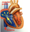
  1. positive jugular pulse, venous distension
  2. ventral edema
  3. signs of right sided heart failure (tachycardia, ventral edema)
30
Q

what pathology can severe tricuspid insufficiency lead to

A

leads to regurgitation of blood into right atrial –> right atrial dilation –> jugular pulsation and peripheral edema

31
Q

what can tricuspid insufficiency be secondary to

A

severe tricuspid insufficiency may be secondary to pulmonary hypertension –> mitral valve regurgitation and increased preload waiting to return to left side –> eventually contributes to secondary pulmonary hypertension which can cause tricuspid regurgitation

scrutinize for concurrent left sided disease or pulmonary disease

32
Q

what type of murmur will a ventricular septal defect produce and where is the PMI

A

grade 4/6-5/6 band shaped systolic murmur

PMI: right cranioventral thorax

33
Q

is S1 and S2 audible in a ventral septal defect murmur

A

no it obliterates S1 and S2

wide radiation and thrill

34
Q

what type of murmur is similar to ventricular septal defect

A

similar murmur heard on left base with PMI over pulmonic valve

caused by relative pulmonic stenosis due to L-R shunt

if not audible on left, tricuspid valve regurgitation is more likely

35
Q

what side of the thorax is a ventricular septal defect heard more loudly

A

on the right but it is detectable on the left as well

36
Q

are horses with ventricular septal defect suitable for athletic use

A

no

37
Q

when could a horse with ventricular septal defect be suitable for athletic use

A

if there is restrictive size

normal RV volume

nromal interventricular septum motion dectected on echo

but unlikely to be suitable top performers

38
Q

when will a horse with ventricular septal defect likely start to decompensate

A

usually 3-4 years

39
Q

what is shown here

A

ventricular septal defect

40
Q

what are the risk factors for ventricular septal defect

A
  1. young animals
  2. arab foals
  3. welsh ponies
41
Q

how is ventricular septal defect diagnosed

A

echocardiography

shows an LV outflow tract view and lesions beneath aortic valve leaflets

42
Q

what size of ventricular septal defect can horses live a normal life

A

if its less than 5mm

43
Q

what are good prognositc indicators of ventricular septal defect

A
  1. small defect (0.5-2.5cm)
  2. high velocity jet of >4.5 m/s
  3. no significant increase in LV dimension
  4. no significant concurrent aortic or mitral regurgitation
44
Q

what are poor prognostic indicators of ventricular septal defect

A
  1. large defect >2.5cm
  2. low velocity jet (higher volume)
  3. increase in LA and LV dimension
  4. significant concurrent aortic or mitral regurgitation
45
Q

what is tetralogy of fallot

A
  1. over-riding right sided aorta
  2. pulmonic artery stenosis
  3. ventricular septal defect
  4. RV hypertrophy
46
Q

what type of murmur occurs with tetralogy of fallot

A

left sided systolic murmur > grade 3/6

47
Q

how is tetralogy of fallot diagnosed

A
  1. cyanosis and poor growth
  2. R-L shunting across VSD because of RV outflow obstruction (stenotic PA)
  3. left sided systolic murmur > grade 3/6
48
Q

what type of murmur is aortic insufficiency

A

grade 2/6-4/6 holodiastolic murmur

49
Q

where is the PMI of aortic insufficiency

A

left base over aortic valve radiating to left cardiac apex

50
Q

what is the signalment of aortic insufficiency

A

usually in older horses with degenerative changes to the valve

51
Q

what should you also do when grading an aortic insufficiency murmur

A

measure the HR

resting HR >40 bpm may be significant

also palpate the pulse pressure –> bounding or weak

52
Q

why is it important to palpate the pulse pressure in aortic insufficiency

A

if it’s bounding the murmur is likely significant

but if the horse is decompensating then it may be weak

53
Q

what can aortic aortic insufficiency lead to

A

left sided overload can lead to pulmonary hypertension which can cause right sided failure –> exercise intolerance and if horse is decompensating then it will have a rapid heart rate and decrease the oxygen getting to the heart and cause fatal dysrhythmia –> potential for acute collapse

54
Q

what is more likely to cause right sided failure in horses

A

left sided overload

55
Q

what predisposes a horse to atrial fibrillation

A

atrial enlargement that occurs with significant aortic or mitral insufficiency

56
Q

what are poor prognosis indicators of aortic insufficiency (6)

A
  1. weak or bounding pulse
  2. LV overload and hyperkenesis
  3. valve thickening
  4. enlargement of aortic root
  5. concurrent ventricular dysrhythmia
  6. concurrent mitral regurgitation
57
Q

what does severe aortic regurgitation lead to

A

left ventricle enlargement

58
Q

how do you assess the severity of aortic regurgitation

A
  1. area of regurgitant jet and velocity
  2. size of left ventricle
  3. pulmonary hypertension and PA distention
  4. exercising ECG for presence of ectopic beats
  5. any mitral valve regurgitation
  6. measure size of aortic route
59
Q

what is the significance of aortic insuficiency

A

significance in young animals

grades > 2/6 need careful evaluation

rate of progression must be assessed

may be extreme tachycardia and poor ventricular bloodflow at higher levels of exercise

acute cardiac output failure at exercise may lead to acute collapse

60
Q

where is the PMI of pulmonic insufficiency

A

rarely able to auscultate

very unlikely to cause cardiac compromise

61
Q

how is pulmonic insufficiency diagnosed

A

echocardiography

62
Q

what is the pathogenesis of bacterial endocarditis

A
  1. damage to endothelium/valve substance
  2. platelet adhesion and fibrin formation
  3. bacterial colonization
63
Q

what bacteria can cause bacterial endocarditis

A

Streptococcus or Actinobacillus

64
Q

what valves are most commonly affected in bacterial endocarditis

A

aortic > mitral > tricuspid > pulmonary valve

65
Q

what are the clinical signs of bacterial endocarditis (7)

A
  1. intermittent fever
  2. murmur
  3. weight loss
  4. lethargy
  5. depression
  6. secondary septic arthritis
  7. renal or pulmonary complications
66
Q

how is bacterial endocarditis diagnosed

A
  1. echo
  2. blood culture when febrile
  3. inflammatory leukogram and raised acute phase proteins
67
Q

how is bacterial endocarditis treated

A

gentamicin (6.6 mg/kg QID) and penicillin G (50,000 IU/kg QID IV) prior to bacterial culture results

68
Q

what is the prognosis of bacterial endocarditis

A

guarded

persistant valvular incompetence

69
Q

what are the causes of pericardial effusion and pericarditis

A
  1. septic
  2. non-septic
  3. neoplastic
  4. idiopathic
70
Q

what might pericardial effusion be a sequel to

A

pleuropneumonia

EHV-1

or part of generalized congestive heart failure

71
Q

how does pericardial effusion and pericarditis lead to cardiac insufficiency

A
  1. fluid accumulation and thickened/inflammed pericardium
  2. cardiac tamponade
  3. decreased diastolic filling
  4. cardiac insufficiency
72
Q

what other tests could be used to diagnose murmurs

A
  1. blood work
  2. PaO2
  3. PaCO2
  4. pH
73
Q

what blood work indicators can assist with murmur diagnosis

A
  1. chemistries: may reflect hypoperfusion of organs if heart failure (prerenal azotemia, increased liver enzymes, lactic acidosis)
  2. serum cardiac troponin I may be increased with myocarditis
  3. white blood cell counts informative if any inflammatory response (left shift neutrophilia)
  4. increased fibrinogen and in serum A amyloid in endocarditis