Congenital Murmurs in Puppies & Kittens Flashcards
what are pathological cardiac murmurs
- incompetent or stenotic valves
- flow through shunts
what are physiological (functional) murmurs
changes in blood viscosity or velocity (anemia), hypoproteinemia, fever
also athletes (big stroke volume)
what are innocent murmurs
soft (max G1-2/6)
systolic, short, variable, localized
young animals
what are not innocent murmurs
NEVER diastolic or continuous
what are causes of murmurs in kittens and puppies (3)
- could be innocent flow murmur
- systemic disease (anemia)
- could be acquired disease but very unusal (early onset cardiomyopathy in cats, myocarditis in puppies)
what are almost all louder systolic mumurs and all continuous murmurs due to
congenital anatomical defects
what are the characteristics of innocent murmurs (3)
- low grade systolic murmur
- left heart base
- usually gone by 16-20 weeks
if G3+ most likely congenital disease (not innocent)
what are the causes of congenital murmurs in dogs in order of prevelance (8)
- aortic stenosis
- patent ductus arteriosus
- pulmonary stenosis
- ventricular septal defect
- mitral valve dysplasia
- tricuspid valve dysplasia
- tetralogy of fallot
- persistent right aortic arch
what are the causes of congenital murmurs in cats in order of prevelance (6)
- ventricular septal defect
- mitral valve dysplasia
- tricuspid valve dysplasia
- aortic stenosis
- persistent right aortic arch
- tetralogy of fallot
what are the causes of congenital murmurs
majority is unknown
some may be genetic (known prevalence in pure bred dogs; some screening in some breeds ex boxer)
which breed has a genetic predisposition to aortic stenosis
newfoundland
autosomal dominant with modifiers
what breeds have a genetic predisposition to patent ductus arteriosus
poodle
polygenic
female:male 2-4:1
what breeds have a predisposition to tetralogy of fallot
keeshond
polygenic
what breeds have a predisposition to pulmonary stenosis
beagle
polygenic
what breeds have a predisposition to persistent right aortic arch
german shepherd
polygenic
how are congenital murmurs diagnosed (7)
- history
- breed/sex/age
- physical exam
- thoracic radiography
- echocardiography
- electrocardiography
- angiography
what are left base murmurs and when would they be heard (4)
- patent ductus arteriosus (continuous)
- aortic stenosis (systolic)
- pulmonary stenosis (systolic)
- innocent/functional murmurs (systolic)
what murmurs can be heard at the left apex and when can they be heard
- mitral valve dysplasia (systolic)
hat are right side murmurs and when can they be heard (3)
- tricuspid valve dysplasia (systolic)
- ventricular septal defect (systolic)
- tetralogy of fallot (systolic, also at left base)
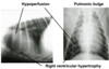
what will thoracic radiography show (3)
- chamber enlargement: left or right sided
- pulmonary circulation: vascular congestion or decreased vascularity
- great vessel dilation
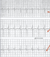
what will electrocardiography show
- left sided enalrgement: aortic stenosis, PDA
- right sied enlargement: right axis deviation –> pulmonary stenosis, tetralogy of fallot
what will echocardiography show (6)
- chamber dilation
- wall hypertrophy
- abnormal valve appearance
- valvular incompetence
- high velocity flow across valves
- shunts
what is the pathology of aortic stenosis
- sub-aortic stenosis (muscular ridge)
- valvular aortic stenosis (valve cusps or annulus)
turbulent flow across aortic region
muscular or fibromuscular ridge or ring beneath the aortic valve
sometimes there is valvular stenosis where the valve cusps are abnormal
can get secondary changes to the valve due to the subaortic stenosis
what is the pathophysiology of aortic stenosis

what breeds is aortic stenosis common in
Rottweiler
boxer
GSD
newfoundland
golden retriever
is aortic stenosis common in cats
rare
may be supra valvular
what is seen on history in animals with aortic stenosis (5)
- asymptomatic
- exercise intolerance
- syncope
- sudden death (can be first clinical sign)
- left sided congestive heart failure (dyspnea, tachypnea, cough)
what is seen on clinical exam with aortic stenosis (3)
- weak pulse
- harsh systolic murmur left heart base
- +/- left sided CHF
what is seen on radiography with aortic stenosis
- cardiac silhouette may be elongated
- post valvular dilation of aorta may be present

what is seen in ECG with aortic stenosis (3)
- tall R waves (left ventricular hypertrophy)
- ventricular premature complexes
- ST segment depression (myocardial ischemia)

what is seen on echocardiography with aortic stenosis (5)
- left ventricular concentric hypertrophy
- aortic valve dysplasia
- sub valvular narrowing of aorta
- high velocity flow across aorta
- turbulent blood flow across aorta

what is the therapy and prognosis of aortic stenosis
mild cases: no treatment, good prognosis
severe: beta-blockers (reduce HR, reduce outflow gradient, reduce risk of arrhythmias and sudden death), calcium channel blockers (diltiazem)
guarded prognosis: little/no evidence that beta-blockers, balloon dilation or surgery help, a cutting ballon may be more effective
increased risk of bacterial endocarditis
what does aortic stenosis increase the risk of
increased risk of bacterial endocarditis
what is patent ductus artiosus (PDA) pathophysiology
left to right shut causing volume overload of lungs and of the left heart
depending of the size of the ductus may progress quickly (1-3 years) to left sided CHF so early closure is recommended
what can also occur in PDA pathophysiology
pulmonary hypertension –> right to left shunt –> caudal cyanosis
what breeds can be affected by PDA
collie
sheltie
CKCS
toy poodle
GSD
spaniel
what is seen on history with PDA (2)
- exercise intolerance
- left sided CHF (dyspnea, tachypnea, cough)
what is seen on clinical exam with PDA
- continuous murmur left heart base
- water-hammer pulse also called machinery murmur
what is seen on thoracic radiography with PDA (3)
- over perfusion of lungs
- +/- pulmonary edema
- aortic, pulmonary and left auricular bulge on DV view

what is seen on echo with PDA (3)
- turbulent flow dlow in pulmonary artery
- ductus visible joining pulmonary artery
- continuous high
what is seen on ECG with PDA
- tall R waves (LV enlargement)

what is the treatment of PDA (2)
- surgical ligation or coil/amplatz plug implantation
- treat left sided CHF if present with diuretics and ACE inhibitors
what is reversed PDA
- excessive volume overload of lungs may lead to pulmonary hypertension (PH) usually occurs early in life (large ductus) but may also occur later complication
- right to left (sometimes bidirectional) shunting into descending aorta occurs due to pulmonary hypertension
what clinical signs occur with reversed PDA
- hindquarter weakness/caudal cyanosis
- may develop polycythemia due to hypoxia
what is the treatment of reversed PDA
treat polycythemia by phlebotomy +/- hydroxycarbamide
what is the prognosis of reversed PDA
guarded/poor
but if respond to sildenafil for PH and management of polycythemia may do okay
what is the pathology pulmonary stenosis
- valvular: dusion, thickening, hypoplasia
- subvalvular: usually due to secondary hypertrophy

what might cause pulmonary stenosis
may be associated with aberrant coronary anatomy in bulldogs and other brachycephalics
what is the pathophysiology of pulmonary stenosis

what are the breeds that are affected by pulmonary stenosis
west highland terrier
bulldog
boxer
cocker spaniel
beagle
is pulmonary stenosis common in cats
no
what is seen on history with pulmonary stenosis (5)
- asymptomatic
- exercise intolerance
- syncope
- sudden death
- right sided CHF (ascites, dyspnea)
what is seen on the clinical exam with pulmonary stenosis
- weak pulse
- harsh systolic left heart base murmur
- ascites
what is seen on radiography with pulmonary stenosis (3)
- hypoperfusion
- pulmonic bulge
- right ventricular hypertrophy

what is seen on ECG with pulmonary stenosis
deep S waves in leads I, II, III

what is seen on echocardiography with pulmonary stenosis
- thickening/fusoin of pulmonary valve +/- valve hypoplasia
- right ventricular hypertrophy

how are mild cases of pulmonary stenosis treated
no treatment
good prognosis
how are severe cases of pulmonary stenosis treated
- beta blockers
- balloon valvuloplasty (works best if fused vavles)
- surgical including patch graft
guarded prognosis
what is the pathophysiology of ventricular septal defect
small perimembranous occurs in the portion of the interventricular septum beneath the aortic valve in the outflow segment of the left ventricle

what breeds are effected by ventricular septal defects
bulldog
keeshond
cocker spaniel
and common in cats
what is seen on history with ventricular septal defect
- asymptomatic
- exercise intolerance
- left sided congestive heart failure (dyspnea, cough)
what is seen on clinical exam with ventricular septal defect
- systolic murmur right ventral thorax (usually) –> the smaller the VSD, the louder the murmur
- good pulse
what are the signs of tetralogy of fallot (4)
- collapse
- dyspnea
- cyanosis
- exercise intolerance
what is seen on the physical exam with tetralogy of fallot (3)
- +/- murmur depends on presence of polycythemia
- +/- cyanosis
- +/- dyspnea
what is the pathology of tetralogy of fallot (4)
abnormal spinal septum formation
abnormality formation of the spiral septum which is in the embryo divides the trunkus arteriosus into the aorta and pulmonary –> deviated to the right which leaves a large VSD
- pulmonary stenosis
- large VSD
- over-riding aorta
- right ventricular hypertrophy
what is mitral valve dysplasia
insufficiency only OR stenosis and insufficiency
how does mitral valve dysplasia present
systolic murmur over mitral valve but if stenosis present diastolic murmur also present
may develop left sided CHF, +/- arrhythmias
how is mitral valve dysplasia treated and what is the prognosis
similar to mitral valve endocardiosis
prognosis depends on severity
what is seen on echo with mitral valve dysplasia
dysplastic valve
leaking valve
how does tricuspid valve dysplasia present (8)
- asymptomatic
- exercise intolerance
- syncope
- may develop rapid supraventricular tachycardia
- +/- ascites
- systolic murmur over right apex
- +/- signs of right sided CHF
- +/- episodic weakness/tachypnea if arrythmia
how is tricuspid valve dysplasia treated (3)
- treat CHF as usual
- surgery (replacement/repair) has been performed in dogs
- manage arrhythmia if present: medical or ablation of accessory pathway