Acquired Murmurs in Adult Dogs Flashcards
what are endocardial diseases of acquired heart murmurs in dogs
- degenerative (mitral) valve disease (DMVD), myxomatous (mitral) valve disease (MMVD), endocardiosis
- endocarditis (usually bacterial)
what are the myocardial causes of acquired heart murmurs in dogs (4)
- dilated cardiomyopathy (primary, secondary)
- arrhythmogenic right ventricular cardiomyopathy
- hypertrophic cardiomyopathy (secondary) - rare
- myocarditis
what breed is degenerative mitral valve disease/myxomatous mitral valve disease/endocardiosis commonly seen in
CKCS
what is degenerative mitral valve disease/myxomatous mitral valve disease/endocardiosis
myxomatous degenerative of the heart valves and chordae
collagen loss and thickening of the valve margins and chordae leads to progressive distortion of the valve leaflets and the possibility of chord rupture

what is the most common cardiac disease in dogs
mitral valve disease
causes of 75% of all congestive heart failure in dogs
what is the pathophysiology of mitral valve disease

what are the clinical features of mitral valve disease (6)
- common in middle aged (5 years+) or older
- small to medium sized
- pure and mixed breed dogs
- some large breeds
- CKCS, poodles, terriers, dachshunds, chuhuahuas, sight hounds, collies, setters, GSDs
- males > females
what is seen on history with mitral valve disease (4)
- asymptomatic
- signs of left sided cardiomegaly (cough)
- signs of left sided CHF & reduced output: tachy/dsypnea (pulmonary edema), cough, exerciseintolerance
- +/- signs of right sided CHF: ascites, dyspnea due to pleural effusion
what are the clinical findings of mitral valve disease (6)
- systolic murmur over mitral +/- tricuspid valve
- murmur grade +/- related to severity of disease
- early stages good myocardial function and good pulse quality
- +/- dyspnea
- +/- ascites
- +/- arrhythmias
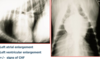
what is seen on chest radiographs with mitral valve disease (2)
- cardiomegaly
- airway compression

what is the cause of the cough in mitral valve disease
main left stem bronchial compression by the enlarged heart (LA) is a relatively common cause of a cough
how does CHF by mitral valve disease show on radiography
- cardiomegaly
- venous congestion
- alveolar pattern

what is shown on the ECG with mitral valve disease (4)
- chamber enlargement: large R wave
- supraventricular premature beats: from atria
- atrial fibrillation
- ventricular arrhythmias

what is seen on echo mitral valve proplapse
enlarged LV and LA
mitral valve cusp prolapsing into LA

what biomarker can be used to diagnose acquired murmurs
NT-proBNP
how is mitral valve disease treated
depends on severity
severity is based on congestive heart failure classification scheme to guide treatment
what is the prognosis of mitral valve disease
gaurded when/if CHF develops 6-24 months survival
more rapid progression in CKCS and large breeds
what is bacterial endocarditis
affects valves on the left side of the heart
which valve is most commonly affected in what is bacterial endocarditis in dogs
aortic valve
which valve is the second most commonly affected in what is bacterial endocarditis in dogs
mitral valve
what predisposes dogs and cats to bacterial endocarditis
congential aortic stenosis
what are the clinical findings of bacterial endocarditis (3)
- pyrexia
- joint pain/stiffness & (shifting) lameness
- new left sided murmur
how is bacterial endocarditis diagnosed (7)
- blood culture
- urine analysis & culture
- +/- joint taps
- echocardiography
- thoracic radiography
- abdominal ultrasonography
- hematology and blood biochemistry
what is the treatment of bacterial endocarditis
- treat with antibiotics (pref based on C&S) often IV to start, supportive therapy as needed
may eventually develop left CHF
prognosis is guarded
how is bacterial endocarditis prevented
treat dogs with severe aortic stenosis with prophylactic antibiotics if undergoing routine dental treatment or if they have wounds
what are the most common myocardial diseases (4)
1. dilated cardiomyopathy (DCM)
2. arrhythmogenic right ventricular cardiomyopathy (ARVC): specific breeds
rare:
- hypertrophic cardiomyopathy (secondary)
- myocarditis (probably under-diagnosed)
what is the etiology of primary dilated cardiomyopathy in dogs
- idiopathic
- hereditary/familial (dobermann, boxer)
what is the etiology of secondary dilated cardiomyopathy in dogs (3)
- toxic: doxorubicin
- tachycardia induced
- dietary: taurine, carnitine deficiency, grain free fad diets
what is the pathophysiology of DCM

what the predispositions of DCM in dogs (3)
- medium/large breeds (overlap with MMVD)
- usually middle age/odler
- males > females
what is seen on history with DCM (5)
- forward failure: exercise intolerance
- left sided CHF: cough, dyspnea, tachypnea, exercise intolerance
- right sided CHF: pleural effusion, ascites
- syncope: ventricular arrhythmia (boxer, dobermann, great dane)
- asymptomatic: occult or early form
what are the physical findings of DCM (6)
- forward failure: pallor, poor CRT, weak pulse, tachycardia
- backward (congestive) heart failure: tachypnea/hyperpnea/dyspnea –> left sided CHF
- hepatomegaly, ascites –> right sided CHF
- arrhythmias
- gallop sounds
- low grade systolic murmursover mitral +/- tricuspid valves
what is seen on radiography in DCM
- left atrial enlargement
- left ventricular enlargement
- +/- signs of CHF
- right sided CHF causing pleural effusion

what is seen here
right sided CHF causing pleural effusion caused by DCM

what is seen on ECG with DCM (3)
chamber enlargement and arrhythmias
- left atrial and ventricular enlargement
- ventricular arrhythmias
- atrial fibrillation

what is seen on echocardiography in DCM
- poor systolic function –> dilated poorly contracting LV
- mitral valve insufficiency –> regurgitation secondary to the distortion of LV
- LA enlargement
how is DCM treated (5)
- improve systolic function
- decrease preload
- decrease afterload
- treat arrhythmias
- reduce further remodelling
how is systolic function improved in DCM (3)
- pimobendan
- digoxin
- dobutamine (IV acute/collapsed cases)
how is preload decreased in DCM (2)
- diuretics (furosemide, torasemide, spironolactone)
- ACE inhibtors (benazepril)
how is afterload decreased in DCM (2)
- ACE inhibitors
- inodilator (pimobendan)
how are arrhythmias treated in DCM (2)
- atrial fibrillation: digoxin, calcium channel antagonists (diltiazem), beta-blockers (atenolol, sotalol, with care)
- ventricular arrhythmias: mexiletine, beta-blockers (atenolol), potassium channel blockers such as sotalol
how is remodelling reduced in DCM (3)
- ACE inhibitors
- pimobendan
- spironolactone
what is the prognosis of DCM
progressive disease
prognosis differs between breeds: survival short in dobermann, sudden death in great danes
6-18 months after onset of CHF
what is occult dilated cardiomyopathy in dogs
DCM may be present in a hidden or “occult” form for several years before CHF develops
what breeds are predisposed to occult DCM
- irish wolfhounds, newfoundlands –> may show slow atrial fibrillation
- boxers, doberman pinschers may just have ventricular arrhythmias
how is occult DCM managed (2)
- arrhythmias: treat life threatening ventricular arrhythmias (sotalol, mexiletine)
- arryhthymias: treat fast atrial fibrillation >160bpm (digoxin, diltiazem, atenolol)
what is boxer cardiomyopathy
arrhythmogenic right ventricular cardiomyopathy
how does arrhythmogenic right ventricular cardiomyopathy present in boxers and bulldogs
in boxers: familial often late onset
autosomal dominant with reduced penetrance
what are the signs of arrhythmogenic right ventricular cardiomyopathy (3)
- ventricular arryhthmias mainly
- poor RV systolic function +/- later LV (“DCM”)
- right sided cardiomegaly initially
what is the pathophysiology of arrhythmogenic right ventricular cardiomyopathy (3)
- arrhythmias: mostly tachyarrhythmias, sometimes bradyarrhythmias –> syncope (forward failure)
- poor myocardial function: pulmonary edema and ascites (left and right sided congestive heart failure backward failure)
- rapid ventricular tachycardia: cardiogenic shock
what is the fatty infiltrative form of arrhythmogenic right ventricular cardiomyopathy and what breed is it seen in arrhythmogenic right ventricular cardiomyopathy
myocytes are replaced with fibrofatty cells

how is arrhythmogenic right ventricular cardiomyopathy managed (3)
- support systolic function with pimobendan
- reduce futher remodelling with ACE inhibitors/spironolactone
- treat symptomatic/life threatening ventricular arrhythmias with anti-arrhythmic drugs such as sotalol/mexiletine/atenolol/amiodarone
what are the criteria for treatment of ventricular arrhythmias (3)
- runs of venticular tachycardia >180bpm
- mutiforms of ventricular premature complexes
- close coupling of ventricular ectopics: R wave on previous T wave

what are secondary cardiomyopathies in dogs (2)
- hypertrophic cardiomyopathy may be seen secondary to systemic hypertension (treat hypertension)
- poor systolic function may be seen secondary to hypothyroidism, doxorubicin therapy (treat endocrine disease, monitor cardiac function on doxo therapy, modify diet if required)
what are the causes of myocarditis in dogs and cats (6)
may be secondary to
- toxoplasmosis
- neospora
- parvovirus
- erhlichia
- borrelia (lyme disease)
- neoplasia
what are the history and clinical findings of myocarditis in dogs and cats (4)
not specific for cardiac disease
- arrhythmias common
- pyrexia
- poor systolic function and secondary dilation with low grade murmurs
- may develop CHF
how is myocarditis diagnosed
difficult but high serum toponin I is supportive of diagnosis
myocardial biopsy
how is myocarditis treated
- treat primary disease if identified
- treat arrhythmias
- treat CHF if present
what is the prognosis of myocarditis
guarded
sudden cardiac death common