ECG - Rhythm strip Flashcards
Learning objectives
- To understand the basic physiology involved in electrocardiography
- To understand and utilise a stepwise approach to ECG rhythm interpretation
Conduction and its problems
Normal depolarisation should begin in the SA node
This conduction can be delayed or ‘blocked’ at any point in the pathway:
- SA node
- AV node
- His bundle
- Bundle branches
Rhythm is best interpreted in lead II or V1 (P wave most clearly seen)
AV node
- PR interval = SA node ventricular muscle
- Normally <220 ms (six small squares)
- If process abnormal it is normally called a ‘heart block’
Arrhythmias
Blocks:
- AV
- Bundle
Brady:
- Sinus
- Siniu node dysfunction
Tachycardia:
Narrow:
- regular
- irregular
Wide:
Bradycardias
- Bradycardias (<60bpm), usually caused by diseases affecting sino/atrioventricular nodes or the conducting tissues of the heart
- Can be normal physiological states
- Need for treatment based on haemodynamic state of the arrhythmia. Not ECG
First degree heart block
If delay between SA ventricles = PR interval prolongation
= ‘First degree’ heart block
Normally causes no symptoms
Can be a sign of:
- CAD
- Rheumatic carditis
- Digoxin toxicity
- Electrolyte disturbances

Second degree heart block
This occurs when excitation ‘misses’ the AV node and bundle of His
Mobitz type 1 (Wenckenbach)
PR gradually lengthens until there is a dropped beat, then a neat with a short PR, which then gradually lengthens until a further dropped beat etc….
Second degree heart block
This occurs when excitation ‘misses’ the AV node and bundle of His
Mobitz type 2
- Stable PR with occasional dropped QRS (atrial depolarization without ventricular depolarization)
- May be alternate conducted or non-conducted beats (or 2, 3 or 4 non-conducted beats so more P waves than QRS)
- This is called 2:1, 3:1. 4:1 conduction

Third degree heart block
- This occurs when atrial contraction is normal but no beats are conducted to the ventricles
- = The atria and ventricles aren’t ‘talking’
- The ventricles are then ‘instructed’ from a slow escape mechanism (from within the ventricular muscle)
- It is a bradycardiac rhythm so a long strip is normally needed
- No consitency in PR interval
- P waves are regular
- QRS are regular (normally look funny as from an alternate ‘focus’)
*

Tachycardia
- Ventricular rate is >100 bpm = tachycardia?
- The next question to consider is whether this is a broad complex or narrow complex tachycardia?
- A narrow complex tachycardia is usually due to an arrhythmia arising in the atria or the junctional region.
- The exception to this rule of thumb is if there is a co-existing bundle branch block which will cause the ventricular complexes to be wider.
- Broad complex tachycardias usually arise from a focus below the atrioventricular node; in the ventricles
Narrow complex tachycardia
- Sinus tachycardia
- A fib
- A flutter

Broad-complex tachycardia
- VT
- VF
- SVT with BBB

How do we read a rhythm strip?
Having a structural approach is the key!
Don’t panic!
Is it the right patient?
Is there more than one strip?
Grab a piece of scrap paper!

Is there electrical activity?
No?
Check leads and electrodes
If it’s a completely straight line; think twice!
Look at the patient!
Is there a pulse?
If not this is asystole
Yes?
Next step!
What are the ventricles doing?
(QRS rate)
Normal is:
- 60-100bpm
Bradycardia is:
- <60 bpm
Tachycardia is:
- >100 bpm
How to estimate HR?
Count the number of QRS in 6s (30 large squares) and times by 10
Or count the number is 3s (15 large squares) and times by 20
Is it regular or irregular?
(QRS rhythm)
The faster it is the harder it is to see!
Is it totally irregular?
Is there a recurring cycle of variation?
Is it almost regular but with the odd funny beat?

‘Funny-looking beats’(1)
A regular rhythm can look irregular due to ectopics
- Morphology of the QRS is key
- venticlesThey can arise from the atria or the
- If the QRS is narrow (<0.12s) likely to ‘come from’ above the ventricles
- If it is wide (>0.12s) likely ventricular in origin or supraventricular with bundle branch block

Funny looking beats (2)
- If they happen before the next sinus beat was due to occur they are called ‘premature beats’
- if its after a long pause they are called an ‘escape beat’
- They can occur in pairs = couplets
- In threes = triplets
- When they happen alternately with a ‘normal’ beat for a sustained period = bigeminy


What’s the QRS width?
Normal is 0.12s (3 small squares)
If it’s less than this = rhythm is from above the bifurcation of the bundle of His
If it’s more than this = rhythm is from the ventricular myocardium or is associated with bundle branch block
Is there atrial activity
It is difficult!
Don’t guess! You must be sure
P waves can be positive/negative/biphasic
They can be hidden in QRS/ST segments/T waves
A 12 lead can help. V1 and lead II are best
Look at rate and regularity in the same was as the QRS
Are the atria and ventricles talking?

If the interval between the P and the QRS is constant it means the conduction from atria to ventricles is intact
Look at a long strip where possible
Is there evidence of heart block?
Is it flutter with 2:1, 3:1 etc conduction?

Normal Sinus Rhythm

Asystole

P wave asystole
Sinus bradycardia

Atrial flutter with 4:1 block

Atrial flutter 2:1, 3:1 and 4:1

Atrial fibrillation

Ventricular tachycardia

Ventricular fibrillation

Fine ventricular fibrillation

Torsade de pointe

Ventricular ectopies

Bigeminy

Paced rhythm

First-degree heart block

PR interval > 200ms (five small squares)
‘Marked’ first degree block if PR interval > 300ms
Complete heart block
- In complete heart block, there is complete
absence of AV conduction – none of the supraventricular impulses are conducted to the ventricles. - Perfusing rhythm is maintained by a junctional or ventricular escape rhythm. Alternatively, the patient may suffer ventricular standstill leading to syncope (if self-terminating) or sudden cardiac death (if prolonged).
- Typically the patient will have severe bradycardia with independent atrial and ventricular rates, i.e. AV dissociation.

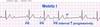
Mobitz type 1

Progressive prolongation of the PR interval culminating in a non-conducted P wave
The PR interval is longest immediately before the dropped beat
The PR interval is shortest immediately after the dropped beat
Mobitz type 1

Progressive prolongation of the PR interval culminating in a non-conducted P wave
The PR interval is longest immediately before the dropped beat
The PR interval is shortest immediately after the dropped beat
