DPT 633: Human Imaging in PT Practice II Flashcards
(58 cards)
Label the following as either permeative, greographic, or moth-eaten

a. Geographic
b. Moth Eaten
c. Permative

Osteopenia

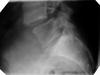
Tear-Drop Fracture

DDD

Os Odontoideum

Scoliosis
Facet Joint Dislocation

DISH

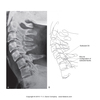
Clay Shoveler’s Fracture

Compression Fracture
Match the following pathologies with the correct radiologic finding
- Spondylosis deformans: _________
- DISH: _________
- DJD: _________
- DDD: _______
- Preservation of disc height
- Decreased zygapophyseal joint space
- Claw-like spurs cupping towards IVD
- Schmorl’s nodes
- Spondylosis deformans: Claw-like spurs cupping towards IVD
- DISH: Preservation of disc height
- DJD: decreased zygapophyseal joint space
- DDD: Schmorl’s nodes
True or False
Typically an old compression fracture (>2months) demonstrates a linear zone of impaction
False
Generalized osteoporosis anywhere in skeleton demonstrates classic radiologic hallmarks of:
a. Trabecular changes
b. decreased radiolucency
c. corticol thickening
d. all of the above
e. none of the above
A
Please match the following decision rules with the correct corresponding predictor variable.
- NEXUS: ________
- Ottawa Ankle Rule: __________
- Canadian C Spine Rule: __________
- Dangerous mechanism of injury
- Tenderness at posterior aspect of tip of lateral malleolus
- No focal neurological deficit
- NEXUS: No focal neurological deficit
- Ottawa Ankle Rule: Tenderness at posterior aspect of tip of lateral malleolus
- Canadian C Spine Rule: Dangerous mechanism of injury

Degenerative Joint Disease

Jefferson’s Fracture

Wedge Fracture
Assuming a T2-weighted MRI, please match the following tissues with the correct signal intensity
- Fluid (CSF, Synovial)
- Fat/yellow marrow
- Cortical bone
- Ligament tendon
- Fluid (CSF, Synovial): High
- Fat/yellow marrow: Intermediate
- Cortical bone: Very low
- Ligament tendon: Low
Factors known to degrade CT image quality include:
a. beam “hardening”
b. metallic implants
c. patient movements
d. all of the above
e. none of the above
D
Please match the following descriptions of Risser’s sign of skeletal maturity with the correct numerical value:
- Osseous fusion is complete
- 100% of crest is “capped”
- 50% of crest is “capped”
- Osseous fusion is complete: +5
- 100% of crest is “capped”: +4
- 50% of crest is “capped”: +2
Please match the following pathologies with the imaging modality of choice.
- Avascular necrosis
- Spinal DJD
- Complex spinal fracture
- Rotator Cuff Tear
- Avascular necrosis: MRI
- Spinal DJD: CT
- Complex spinal fracture: CT
- Rotator Cuff Tear: MRI
True or False
Plain film radiography is the best imaging modality for identifying subtle fractures and/or complex fractures
False
True or False
A Chondrosarcoma is classified as a primary benign bone tumor
False
True or False
The spine is a common region for clinical presentation of a chordoma
True