Day 4, Lecture 2: Genetics V: Human Chromosome Abnormalities Flashcards
Clinical Cytogenetics
- Study of the relationship of chromosomal alterations and genetic diseases in human beings
Chromosomal abnormalities cause
- Pregnancy loss
- Congenital malformations
- Intellectual disabilities (Mental Retardation)
- Play a role in the pathogenesis of malignancy
How common are chromosome abnormalities
- about 1% of live births
- about 50% of first trimester miscarriages
Classification of cytogenetic mutations

Numerical Chromosome Abnormalities

What is the most common chromosome abnormality seen in live births?
- Trisomy 21
- Incidence 1 out of every 691 live births
- Can happen to women of all ages, races and economic levels
Causes of Trisomy 21
- Caused by having 3 compies of chromsome 21
- 95% is nonfamilial Trisomy 21
- 3-4% is unbalanced translation
- only 1% are familial unbalanced translocation
- 1-2% is mosaicism with two cell lines- normal and trisomy 21
Common Aneuploidy Conditions
- Autosomes
- Trisomy 21
- Trisomy 18
- Trisomy 13
- Sex Chromosomes
- Monosomy X
- Klinefelter Syndrome
Clinical Features of Trisomy 21

What is Mosaicism?
- The situation in which not all cells in an organism possess the same genotype. Mosaicism usually occurs as the result of postconceptional (acquired) changes.
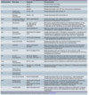
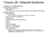
Trisomy 18 (Edwards Syndrome)
Trisomy 13 (Patau Syndrome)

Monosmy X (Turner Syndrome)

Klinefelter Syndrome (47, XXY)

Do Chromosome aneuploidy conditions usually run in families?
- No, it is a sporadic event
Structural Abnormailities

What is an Isochromosome?
One arm is duplicated forming two arms with a shared centromere
Chromosomal Translocations


Microdeletion Syndromes bridge the gap between
- Single gene and chromosomal syndromes
- The deletion of a megabase or so is too small to be seen under the microscope, however, it may involve a dozen or more genes
- FISH analysis and chromosome microarray are revealing increasing numbers of such submicroscopic chromosomal abnormalities
Molecular pathology puts deletions/duplications into what three categories

microdeletion syndromes, contiguous gene syndromes, and segmental aneuploidy are now generally referred to as
- Genome disorders
- (note: Genome disorders have a complex phenotype (frequently includes mental retardation, developmental delay, and behavioral problems) due to alteration of gene dosage)
- Haploinsufficiency
- Deletion (majority of genome disorders)
- Terminal deletion (telomeric)
- Imprinting or UPD
- Overexpression
- Duplication (minority of genome disorders)
- Haploinsufficiency
- (note: Genome disorders have a complex phenotype (frequently includes mental retardation, developmental delay, and behavioral problems) due to alteration of gene dosage)
Mechanism of genome disorders
- Usually less than 5Mb is involved in the deletion or duplication that leads to a genome disorder
- Areas of high recombination are often flanked by unique low copy repeats
- Due to the low copy repeats, the chromatids misalign during recombination in meiosis
- This misalignment leads to non-homologous recombination and unequal crossing-over