Cerebral hemishperes - Grey matter (1st half) Flashcards
Identify the general features of the cerebral hemispheres on the diagram


The elevations in the cerebrum are called Gyri
How is this different from the cerebellum?
In the cerebellum - the bumps are called ‘folia’ (not sure if right spelling)
What are basal ganglia?
Collections of neuronal cell bodies within white matter
They are just CNS ‘Nuclei’
What is this structure here?

Corpus Callosum
Thick band of white matter (nerve fibres) that anatomically divides but neurally connects the two hemispheres of the brain
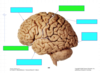
In this photo of the brain, what is the stuff covering the left hemisphere?

Covered in pia mater (the innermost meningeal layer) which is vascularised - hence all the blood vessels
Remember the CSF sits between the arachnoid and pia mater
Identify the fissures in green and the lobes in blue

What is this?

The Pre-central gyrus
The gyrus immediately anterior to the central sulcus
Primary motor cortex
What is this?

Post central gyrus
Primary sensory cortex
Identify the parietooccipital sulcus and central sulcus of the brain in the photo

Parietooccipital sulcus in red
Central sulcus in blue

What gyrus is this arrow pointing to?
What is its associated sulcus?

Cingulate gyrus that runs around the Corpus callosum

What is this area?
Which lobe is it?

The Hippocampus
This is the part of the Temporal lobe that faces medially and is more or less hidden away
Identify the labels boy


If you were to open the lateral sulcus up. You would see this.
What area of the brain is this?

The insula:
The transverse temporal gyri are not on the insular surface. You can see were they are on the brain on the diagram

What is Cortical mapping?
Mapping of 52 areas across the brain, each of which has its own microscopic differences according to its function
These areas are called Brodmann areas
Broadly speaking. The anterior half (frontal lobe) of the cerebrum is associated with _______ functions and the posterior half carries out _______ functions
Frontal lobe (anterior half of brain pretty much) = Motor functions
Posterior (all the others) = Sensory functions
Complete the labels for the general function(s) of each lobe


What is the function of the medial portions of the cerebral hemispheres?
Limbic system - storage and retrieval of processed information
What is meant by primary sensory areas and association areas?
The primary sensory areas are the primary cortical regions of the five sensory systems in the brain. Except for the olfactory system.
So the information received from eg your fingers when you’re touching something goes to your primary sensory area for touch etc
Your association areas take look at the information from your PSA and look for patterns, so you can recognise/appreciate what you’re touching/seeing etc etc
Is there association areas in the frontal lobe? (motor association areas?)
Yeah
They basically formulate the plan (pattern of motor signals) that will achieve a task
So:
- the very front of the frontal lobe will decide ‘im gonna pick up that pen’
- The bit anterior to the primary motor cortex/pre-central gyrus will make the plan for which nerves to fire and when - in order to pick up the pen
- The primary motor cortex will then carry out the instructions made
So thats the job of the motor association areas
What is the primary motor cortex?
Which Brodmann area is it?
Area 4
Somatotopic representation of the contralateral half of the body
This means that all muscles in the other half of the body are represented (controlled) in the primary motor cortex gyrus

What areas of the body require the most space on the primary motor cortex?
Those that carry out fine motor movements
This means the hands, face, tongue require more space than say the hip
What is the pre-motor cortex?
Areas 6 & 8
This is the part which decides how (plans the neurons firing and when) for a motor task
What is the Brocas area of speech?
What Brodmann areas?
Areas 44, 45
These are the areas associated with speech. They are located very close to the areas for hearing as well.

What is the purpose of the pre-frontal cortex?
cognitive functions of higher-order - intellect, judgement, prediction, planning


