Cardiovascular system: Lecture 3 - Development of the Cardiovascular System 1 Flashcards

1
Q

When does the formation of the heart begin?

A

week 3

2
Q

What day is the first heart contraction?

A

Day 22

3
Q

What are the 3 layers that the heart is composed of?

(from inner to outer)

A
  1. Endocardium - derived from the heart tube
  2. Myocardium - derived from the viscerla mesoderm overlying the heart tube
  3. Epicardium - this is the visceral layer of the pericardium and is also derived from the visceral mesoderm
4
Q

What does the myocardium go onto form?

A

Muscular cells that allow contraction of the heart

5
Q

What is vasculogenesis?

A

Making new vessels from scratch

6
Q

Explain how vasculogenesis works?

A
  • the endoderm induces some cells of the overall visceral mesoderm to differentiate into angioblasts
  • angioblasts differentiate into endothelial cells and form endocardial tubes
7
Q

When does embryonic folding begin?

A

Day 17/18

8
Q

What do endocardial tubes form when they fuse during lateral folding?

A

The primitive heart tube

9
Q

What does the myocardium secrete and why is this important?

A

The myocardium secretes cardiac jelly which is important in cardiac looping and septation of the heart

10
Q

How is the developing heart tube brought into the thorax?

A

Craniocaudaul folding

11
Q

What does th endocardium and myocardium go on to form?

A

The internal endothelial lining of the heart and muscular wall respectively

12
Q

What is the cardiac jelly?

A

gelatinous connective tissue separating the myocardium and heart tube endocardium

13
Q

What occurs in the caudaul region during week 4 of embryobic development?

A

3 paired veins drain into the tubular heart via the right and left horn of the sinus venosus: heart is already beating at this point

(blue branches in image)

14
Q

With differential growth of the heart tube, which 5 dilations become apparent?

A

Truncus arteriosus

Conus arteriosus

Ventricle

Atrium

Sinus venosus

These develop into the adult structures of the heart

15
Q

Which direction does blood flow through the heart tube?

A

Bottom to top (caudal to cranial) - from sinous venosus to truncus arteriosus

16
Q

What happens during day 23?

A

The heart tube starts to fold in preparation for dividing into 4 chambers

17
Q

Explain the sequence of events in looping of the heart

A
  1. Straight heart tube
  2. C-shaped loop
  3. S-shaped loop

Bulbus cordis moves caudally, ventrally and to the right

Primitive ventricle is displaced before moving back to midline

Primitive atrium displaces cranially and dorsally

18
Q

When does the sinus venosus degenerate?

A

In week 5

19
Q

What happens to the remnants of the sinus venosus?

A

It remains as part of the wall of the right atrium (right horn) and contributes to the venous drainage (left horn) of the heart

20
Q

What does the left horn of the atrium go on to form?

A

The left horn forms the oblique vein of the left atrium and coronary sinus

21
Q

What does the right horn form?

A

the smooth-walled part of the right atrium – sinus venarum

22
Q

How can the part of the right atrial wall formed from the right horn be distinguished from the rest of the right atrial wall (the majority)?

A

As the rest appears rough - trabeculated and was derived from the primitive atrium

23
Q

What is the crista terminalis?

A

The clear border between the trabeculated part of the right atrium and the sinus vernarum

24
Q

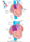
Annotate this image (Name the rough parts and the smooth parts)

A
25
Q

How does the ventricular wall form?

A

Majority by the primitive ventricle, with a small contrbution from the conus arteriosus

26
Q

What does the conus arteriosus form?

A

the smooth walls of the left and right ventricles that lead into the aorta (aortic vestibule) and pulmonary trunk (conus arteriosus) respectively

27
Q

What is the trabeculae carneae?

A

muscular ridges - the rest of the ventricular wall that is trabeculated (rough) and formed from the primitive ventricle

28
Q

Annotated image of the right ventricle

A
29
Q

What does an outgrowth of the left atrial wall form?

A

Initially, it forms a single pulmonary vein. The pulmonary vein branche into left and right veins which then bifurcate to form 4 pulmonary veins

30
Q

What is intussusception?

A

In week 5, the 4 pulmonary veins are incorporated into the wall of the left atrium. This makes the left atrial wall smoother than the right

31
Q

Where is the right sinus horn encorporated into?

A

The right artrium, enlarging it

32
Q

What is the majority of the atrial wall made from?

A

the primitive atrium

33
Q

At the end of week 4, explain how the primitive atrium divides into right and left atria

A

-begins with a crescent-shaped outgrowth from the dorsal wall – septum primum

34
Q

Explain how the atria is seperated from the ventricles

A
  • As the septum primum extends into the atrium, the endothelium lining the boundary between the atrium and ventricle expands to form dorsal and ventral endocardial cushions
  • These cushions fuse in the midline to form the atrioventricular septum with 2 channels communicating between the future left atrium/ventricle and right atrium/ventricle
35
Q

In what week has the septum primum has fused with the atrioventricular septum? What is obliterated?

A

Week 6

The ostium primum is obliterated

36
Q

How do the foramen secundum and the septum secundum form?

A

foramen secundum - Apoptosis occurs in the upper part of the septum primum to form the foramen secundum

seutum secundum - on the right side of the foramen secundum, a second crescent shaped projection forms from the dorsal wall of the atrium – septum secundum

37
Q

What is the opening left in the septum secundum called? Why is this important?

A

the foramen ovale- this allows blood to flow from the right atrium to the left atrium, bypassing the lungs and goes straight to the systemic circulatory system

Foetuses don’t need to send blood to the lungs because the mother provides us with oxygen

38
Q

What is the foramen primum?

A

The diminishing connection between the left and right sides of the primitive atrium as the septum primum extends

39
Q

How is an interventricular foramen formed?

A

At the end of week 4, a muscular part that projects from the floor of the primitive ventricle towards the endocardial cushions, leaving an interventricular foramen

40
Q

When is the intraventricular foramen closed?

A

Week 7

Membranous part that projects inferiorly from the endocardial cushion - seperates ventricles from right and left

41
Q

What does the truncus arteriosus septate into?

A

The aorta and pulmonary trunk

42
Q

How is the truncus arteriosus divided?

A

into two channels by endocardial swellings - cronotruncal ridges

43
Q

What do the conotruncal ridges fuse to?

A

They fuse to form a septum (a conotruncal septum) that separates the outflow of left and right ventricles and fuses with interventricular septum

44
Q

Where are neural crest cells contained?

A

The conotruncal swellings

45
Q

Explain how foetal circulation works

A
  • oxygenated blood enters through umbilical vein
  • some blood enters liver and rest enters ductus venosus to bypass liver and enter IVC
  • blood passes through the foramen ovale to the left atrium, ventricle and aorta
  • blood also travels through the SVC and passes through the ductus arteriosus
46
Q

What happens to the ductus venosus when umbilical circulation stops?

A

It closes and degenerates

Forms the ligamentum venosum in aduls

47
Q

What other foetal strucuture closes?

A

the foramen ovale - fossa ovalis in adults