B3 L37: Fractures of the upper limb and their management Flashcards

1
Q

What is the mechanism of injury for clavicle fractures?

A

Fall onto superolateral aspect shoulder > direct blow to shoulder > FOOSH

2
Q

What are 4 other injuries which can occur with clavicle fractures (associate EXAM QUESTION

A
  1. # scapula
  2. # ribs – pneumothorax/ haemothorax/ pulmonary contusion
  3. vascular injuries
    • Subclavian artery
    • Axillary vessels
  4. brachial plexus injuries
3
Q

What is a traction type injury?

A

head and shoulder go in different directions

4
Q

What are medial clavicle fractures usually a result of (MOI)?

A

of high energy blunt trauma such as MVA, MBA, Ped vs car

90% associated with multi trauma

5
Q

What is the most likely to less likely location that is injured in a clavicle? Why is this the case?

A

Mid shaft > lateral> medial

  • two relatively flat surfaces linked by middle tubular section, area of inherent weakness
6
Q

What is the medical management for a medial clavicle fracture?

A

Medial largely non operative unless posterior displacement that threatens neurovascular structures and skin (risk of injection- complications)

7
Q

What is the medical management for a middle clavicle fracture?

A

Middle largely non operative unless significant angulation or shortening

8
Q

What is the medical management for a lateral clavicle fracture?

A

Lateral depends on whether conoid segment of coracoclavicular ligament is attached to the medial fragment of the clavicle

9
Q

What is the medical management for when a ORIF is needed for a clavicle fracture?

A

ORIF may be indicated if pathological fracture, progressive neurological loss, scapulo- thoracic dissociation, open injury, impending skin disruption

10
Q

The Position of fracture can affect chance of _____ due to ligamentous structure. Give 2 examples.

A

healing

  • Fracture medial to coracoclavicular ligaments
  • Fracture between coraoclavicular ligaments
11
Q

What are methods of immobilisation for a clavicle fracture?

A
  • Large number of binder applications for fracture immobilisation
  • Sling application (eg. figure 8 brace shoulder sling (most common)
12
Q

What are 2 characteristics of operative treatment for clavicle fractures?

A
  1. Plate fixation +/- bone graft preferred method (problems- have screw which loose and burrow to other areas of body (eg. heart))
  2. Intramedullary nail less rotational control (nail pass down long bone)
13
Q

What are the 5 important guidelines to follow for clavicle fractures post op?

A
  1. 3/52 sling
  2. gentle active-assist shoulder F and ER in supine (no pendular- due to displacing force of the weight of the arm))
  3. after 6/52 gentle isometric strengthening with progressive resistance beginning @ 2/12
  4. light lifting 3/12
  5. heavy lifting and contact sports restricted until 6/12
14
Q

What is the mechanism of injury for scapula fractures? List 3 mechanism.

A
  1. High energy direct trauma
  2. Axial loading through fall on outstretched arm
  3. Dislocation of the shoulder can fracture glenoid
15
Q

What is the most likely to less likely location that is injured in a scapula?

A

Body> neck> glenoid > acromion > coracoid

16
Q

What are 4 signs and symptoms of a scapular fracture?

A
  1. Arm held adducted
  2. local tenderness
  3. If displaced scap neck or acromial # -> shoulder appears flattened
  4. deep inspiration - pain from attached muscles
    • pec minor - coracoid #
    • serratus anterior - body #
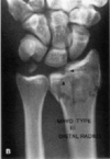
    • with body #’s - there is deep swelling which is often quite painful -> inhibition RC -> loss of arm elevation - resolves in a few weeks
17
Q

What are 8 associated injuries in conjunction with scapula fractures?

A
  1. pneumothorax
  2. rib #’s
  3. pulmonary contusion
  4. # clavicle
  5. brachial plexus injury
  6. arterial injury
  7. skull #’s and CHI (closed head injuries)
  8. spinal #’s
18
Q

What is the complication with an acromial scapula fracture?

A

decrease subacromial space = pressure on structures that pass through

19
Q

What is the management of a glenoid fracture (intra-articular), if it is unstable or if the humeral head is unstable?

A

ORIF and capsular repair

20
Q

What are the 3 types of acromial fractures?

A

Type 1

Type 2

Type 3

21
Q

How is a type 1 acromial fracture classified?

A

minimally displaced

22
Q

How is a type 2 acromial fracture classified?

A

displaced but no reduction in subacromial space

23
Q

How is a type 3 acromial fracture classified?

A

displaced with reduction in subacromial space

24
Q

What are the 2 types of glenoid neck fractures?

A
  1. Without associated separation A/C jt or clavicle #
  2. With above associated injuries
25
Q

What is the management for a glenoid neck fracture without associated separation A/C jt or clavicle #?

A

sling for a few days and then passives

26
Q

What is the management for a glenoid neck fracture with associated injuries?

A
  • may -> discomfort and instability
  • indicating need for surg
  • will also need surgery if angulation is >40 degrees
27
Q

What is the complication of a type 3 acromial fracture?

A

impacting on important structures - acromion moving into subacromial space

28
Q

How should type 1 and 2 acromial fractures be managed? Operative or non-operative? List 3 guidelines to follow?

A
  1. Non-operative sling 3/52
  2. PROM commence Day 1
  3. progress to isometrics and then active movements incorporating RC and deltoid resistance work after # healing
29
Q

How should type 3 acromial fractures be managed? Operative or non-operative? List the guideline to follow?

A
  1. operative treatment - ORIF
  2. immobilization - as cont deltoid contraction could depress acromion preventing healing
30
Q

What are the 2 types of coracoid fractures?

A

Type 1

Type 2

31
Q

How is a type 1 coracoid fracture classified? (2)

A
  1. proximal to C/C (corococlavicular) lig
  2. usually have associated A/C separation, clavicle #, superior scap# or glenoid # and may require Surgical fixation
32
Q

How is a type 1 coracoid fracture classified?

A

distal to C/C (corococlavicular) lig

33
Q

What is the mechanism of injury in a scapula body fracture?

A

High energy blunt trauma or sudden contraction of muscles eg electrocution or seizures Often associated with polytrauma

34
Q

How should a scapula body fracture be managed?

A
  • Mostly conservative management sling and then 1/52 post-injury -
  • pendular and active-assist exercises
35
Q

What is a floating shoulder? What are 2 combined fractures? What 2 directions are they compromised?

A

Combined clavicle (anterior) and scapula (posterior) fracture

36
Q

What is often the management for a floating shoulder?

A

Operative fixation often indicated

37
Q

What is the mechanism of injury of a proximal humeral fracture?

A

Most common is fall on the outstretched hand

38
Q

A proximal humeral fracture is most common in elderly population with__________.

A

osteoporosis

39
Q

A fracture of the proximal humerus occurs in younger persons from ______ such as _______.

A

direct trauma; fall onto the shoulder

40
Q

What are the 3 x rays needed for a shoulder trauma series?

A
  1. True Shoulder AP
  2. Lateral (Scapula Y view)
  3. Axillary view
41
Q

What is a AP shoulder x ray? What are 2 things it can show?

A
  1. Clearly defines the joint space
  2. In absence of subluxation or dislocation should be no overlap between the humeral head and the glenoid
42
Q

What is the true scapula lateral or scapula Y view?

A
  • Right angles to the AP view
  • Beam parallels the scapula spine
43
Q

What can the true scapula lateral or scapula Y view show? How does that differ from an AP view?

A

anterior and posterior dislocation and displaced tuberosities

normally AP can tell if there is displacement but not direction

44
Q

What is the axillary view on the x ray? What position should the arm be in?

A

Arm abducted 20 – 30 degrees

45
Q

What are 3 things the axillary view show?

A
  1. Best for evaluation of articular surfaces of humeral head and glenoid
  2. Fractures of the humeral head
  3. Posterior fracture dislocations and displaced greater tuberosity fractures
46
Q

How does Neers classify proximal humeral fractures? What are the 4 different types?

A
  1. Head of Humerus
  2. Greater Tuberosity
  3. Lesser Tuberosity
  4. Shaft

Part 1

Part 2

Part 3

Part 4

47
Q

How is a fracture classified as displaced (proximal humeral fracture)?

A

>1cm separation or >45 degrees angulation otherwise one part fracture

48
Q

What does a part 2 (SNOH) proximal humeral fracture look like on an x-ray?

A

significant displacement through surgical neck of humerus

49
Q

What does a part 2 (GT) proximal humeral fracture look like on an x-ray?

A
50
Q

What does a part 3 proximal humeral fracture look like on an x-ray?

A
51
Q

What does a part 1 proximal humeral fracture look like on an x-ray?

A

complete

52
Q

How should a 1 part proximal humeral fracture be managed? (list 4 guidelines)

A
  • Most common NOH #
  • Conservative management usually most appropriate (without surgery- which make it worse Part 1 –> part 2)
  • Period of immobilisation in sling (stiff shoulder vs healing of fracture)
  • Passive exercises till union then active
53
Q

How should a 2 part proximal humeral fracture be managed?

A
  • Closed reduction usually initial management (traction force–> fracture back into position )
  • If not able to be reduced or unstable can be fixed with a number of options
  • Percutaneous pins, bone sutures, cancellous screws, intramedullary pins with TBW or locked nail
54
Q

What is a Closed reduction percutaneous fixation?

A
  • low risk of displacement and further complications - high risk of infection (breakage of skin)
55
Q

How should a 3 part proximal humeral fracture be managed? Why?

A
  • Usually displacement of surgical neck and greater tuberosity (increasing velocity = increase displacement)
  • Difficult to reduce closed and usually require ORIF
56
Q

How should a 4 part proximal humeral fracture be managed? Why?

A
  • Surgical neck and both tuberosities
  • Poor outcomes from internal fixation so Hemiarthroplasty usually treatment of choice
57
Q

What are 4 complications of a proximal humeral fracture?

A
  1. Vascular and nerve injuries (axillary)
  2. Glenohumeral stiffness
  3. Malunion (can be reasonably well tolerated in the shoulder)
  4. AVN
    • 10% of 3 part fractures
    • 20% of 4 part fractures
58
Q

It is important liaise with Orthopaedic team in regard to commencing movement of the shoulder (usually commence with passive or pendular exs) during a proximal humeral fracture. What are 6 guidelines to follow post op?

A
  1. Balance between stability of the fracture and prevention of long term stiffness
  2. Mobilisation of adjacent joints
  3. Management of oedema
  4. Cryotherapy
  5. Sling application (well fitting sling -> relax –> pain relief)
  6. Mobility and falls assessment as indicated if stop at UL –> can continue to cause problems (eg. other side
59
Q

What is the capitellum?

A

most distal portion of lateral column

60
Q

What is the trochlea?

A

intermediate between capitellum and distal end of medial column

61
Q

What is the medial epicondyle?

A
  • most distal portion of medial column
  • Closely related to ulna nerve
62
Q

What is the lateral epicondyle?

A

projected forwards from shaft 40 degrees

63
Q

What is the mechanism of injury for a distal humeral fracture?

A

Axial load through flexed >90 degrees

64
Q

How can a distal humeral fracture xray differ?

A

Need traction x-ray so as can see pieces without overlap.

65
Q

Distal humeral fractures __ are/are not common.

A

Are not (when they do occur, they are very severe)

66
Q

What are the 3 ways distal humeral fractures are classified?

A

Type A

Type B

Type C

67
Q

What is a type A distal humeral fracture?

A

Non-articular

68
Q

What is a type B distal humeral fracture?

A

Partially articular – part of articular segment remains in continuity with shaft

69
Q

What is a type C distal humeral fracture?

A

 Articular with no fragments remaining in continuity with shaft  C1 – simple T of Y #  C2 – articular # simple but non-articular supracondylar comminuted  C3 – articular segment comminuted  Prognostically further

70
Q

What is a type C1 distal humeral fracture?

A

simple T of Y #

71
Q

What is a type C2 distal humeral fracture?

A

articular # simple but non-articular supracondylar comminuted

72
Q

What is a type C3 distal humeral fracture?

A

articular segment comminuted

73
Q

What are the 2 operative treatment options (and all the post op management) for distal humeral fractures?

A
  1. ORIF – is the treatment of choice
    • Rehab
      • Immobilised 10/7 and then commence AROM
      • If concerned about stability may then use a ROM brace
      • The aim is to restore anatomy and fix it reliably enough to permit painless early function while bone goes on to uneventful healing.
  2. EF
74
Q

What are the 3 treatment options (and all the management processes) for distal humeral fractures?

A
  1. Traction (3/52) followed by cast brace/hinge brace
  2. Collar and Cuff – in as much elbow flexion as possible to allow gravity to exert a ligamentotaxic effect
  3. Rehab
    • Shoulder pendular at 10 days
    • Elbow movement approximately 3/52
    • Healing usually at 6/52 when collar and cuff discontinued and more specific extension exercises begun.
75
Q

When would non-operative management be chosen in a distal humeral fracture?

A
  • Frail, cardiac CI
  • Open wounds/degloving injuries
  • Extreme OP
76
Q

What are 2 other fractures around the elbow?

A
  1. Olecrannon fracture
  2. Radial Head fracture
77
Q

What are the 2 major principles of management for fractures?

A
  1. Stable fracture
  2. Early ROM
78
Q

What is the mechanism of injury for an olecranon fracture?

A

Either by direct or indirect trauma

  • Direct Trauma: A fall or blunt trauma on the posterior tip of the elbow
  • Indirect Trauma: avulsion of the olecranon by forces generated within the triceps muscle. May occur during a fall onto the partially flexed elbow
79
Q

What is the x-ray view that is crucial for an olecranon fracture? Why? (4)

A

Lateral x-ray

  • Extent of the fracture
  • Degree of comminution (management approach that should be taken)
  • Amount of disruption of art. surface in the semilunar notch
  • Displacement of the radial head
80
Q

What are the 2 classification types of an olecranon fracture?

A

Type I

Type II

81
Q

What is a type I olecranon fracture?

A

Non-displaced and Stable – ie. No change in position with gentle flexion to 90 degrees or extension against gravity

82
Q

What is a type II olecranon fracture? What are the 4 additional classifications of a type II fracture?

A

Displaced Fractures A. Avulsion # B. Transverse/Oblique C. Comminuted # D. Fracture/dislocation

83
Q

What are the 3 guidelines to follow in rehab in a type I (Non-displaced and Stable) olecranon fracture?

A
  1. Immobilised in a long-arm cast in 90 degrees for 2-4 weeks (short period- doesn’t tolerate long period of immobilisation) to allow pain and swelling to diminish.
  2. Followed by protected AROM flexion and passive extension exercises avoiding flexion past 90 degrees until bone healing is complete radiographically (usually 6-8 weeks).
  3. Can then begin passive flexion, active extension and resistive exercises.
84
Q

Should an olecranon fracture be immobilised for a long or short period of time?

A

short- doesn’t tolerate long period of immobilisation

85
Q

What is a type 2(A) olecranon fracture?

A

Avulsion fractures

A transverse or oblique line dislodges a small nonarticular portion of the olecranon, which is displaced by the pull of the triceps muscle

86
Q

What is a type 2(D) olecranon fracture?

A
  • # line usually near the level of the coronoid
  • Ulna will dislocate or subluxate anteriorly, along with the radius
87
Q

What are the 5 operative treatment methods for an olecranon fracture?

A
  1. ORIF with TBW eg avulsion or transverse # (if no comminution)
  2. Intermedullary nail fixation
  3. Combination of IM pin or screw and TBW eg comminuted # or #dislocation
  4. Plate and screws eg. comminuted fracture with bone loss
  5. Excision of the proximal fragment
88
Q

What is tension band wiring?

A

The basic principle is to counteract the tensile forces that act across the fracture site and convert them into compressive forces so that, with motion of the joint, compression across the fracture occurs.

89
Q

How is tension band wiring done?

A
  • Two parallel K-Wires across the fracture site
  • Circlage wire is then passed in a figure of 8 fashion around the insertion of the triceps tendon and then distally beyond the fracture site into a transverse drill hole on the posterior border of the olecranon
90
Q

What is the purpose of tension band wiring?

A

screw- rotatory force –> linear force best environment for

healing loop wire- force of triceps is being transmitted distally (force off fracture to allow healing)

91
Q

What is Post-op TBW Rehab? (4)

A
  1. Within 1 - 2 days gentle active and active assisted motions are started, provided the wound is healing satisfactorily
  2. The elbow can be supported between exercise sessions with a removable splint. Usually support may be discontinued by the 4th week
  3. Similar rehab post IM and Plate & Screw fixation
  4. Maximal function may not return before 6-12months (extensive rehab)
92
Q

What are the 4 disadvantages of non-operative management for displaced olecranon fractures?

A
  1. Healing in elongated position – decreased triceps power
  2. Articular incongruity
  3. Displaced olecranon fragment blocks full extension
  4. Decreased elbow flexion as immobilised in extension to allow bony contact.
93
Q

What are the 3 complications of non-operative management for displaced olecranon fractures?

A
  1. Decreased ROM – up to 50% of cases but some of these not a significant loss
  2. Non-union – 5% of cases
  3. Post-traumatic arthritis – not as common as in WB joints
94
Q

What are 3 characteristics of radial heads?

A
  1. Seated in the lesser sigmoid notch and maintains contact with the ulna throughout forearm pronation/ supination.
  2. With elbow in valgus alignment: main pathway for load transmission is direct axial loading of the radius
  3. In Varus alignment: force transmission is via the radius to the ulna through the interosseous ligament
95
Q

What is the mechanism of injury for radial head fractures?

A
  • Falling on an outstretched hand with the elbow partially flexed and supinated (common)
  • Can also occur by placing excessive axial force on a pronated forearm
96
Q

What are the 4 types of radial head fractures?

A

Type 1

Type 2

Type 3

Type 4

97
Q

What is a type 1 radial head fracture?

A

Undisplaced

98
Q

What is a type 2 radial head fracture?

A

Displaced

99
Q

What is a type 3 radial head fracture?

A

Comminuted

100
Q

What is a type 4 radial head fracture?

A

Fracture Dislocation (10% of radial head fractures assoc with elbow dislocation)

101
Q

What are 2 factors influencing choice of treatment for radial head fractures?

A
  1. High or low demand elbow
  2. Associated Injuries:
    • Interosseous ligament and distal radio-ulna ligament damage
    • Elbow dislocation +/- Coronoid # (gross instability)
102
Q

What are 5 treatment options for a type 1 radial head fracture?

A
  1. Aspirate joint and inject local anaesthetic for pain relief
  2. Patient is given a sling or the arm is splinted for no longer than 3-4 days
  3. Active forearm rotation as soon as tolerated – this is only limited by pain and swelling
  4. Can expect good to excellent function after 2-3 month of active motion exercises
  5. Night time extension splinting if required
103
Q

What are 3 treatment options for a type 2 radial head fracture?

A
  1. Fractures that have no mechanical block: treat as per type 1 injuries with close F/U required and x-rays taken @ 1,2,4 &6 weeks as displacement can occur.
  2. Fractures with mechanical block: ORIF (high demand) vs Excision (low demand).
  3. With Associated Injury: with IOL (Interosseous ligament) tears preservation of the RH is crucial (or RH function with a prosthesis). Excision of the RH may lead to symptomatic migration of the radius. In this situation ORIF of radial head should be strongly considered
104
Q

What are 3 treatment options for a type 3 radial head fracture?

A

Non-repairable

  1. Should undergo early excision as the treatment of choice. Elderly do not require RH replacement and should begin early active motion with passive motion and extension splinting as needed to regain ROM
  2. If radial head requires excision – lateral ligament complex also needs repairing – may need to limit supination initially after this and gradually advance.
  3. If the IOL (interosseous lig) and distal R/U lig is torn then will require metallic radial head arthroplasty to stabilise the forearm.
105
Q

What are 2 Indications for replacing radial head?

A
  1. The Type III unreconstructable radial head # (just discussed)
  2. Elbow dislocation with ligament ruptures +/- coronoid process #
106
Q

What are the 2 x-rays to determine a distal radius fracture?

A
  1. AP
  2. Lateral views to determine deformity:
    • Articular step off
    • Radial shortening
    • Dorsal inclination
    • Radial inclination
107
Q

What is an articular step off in a distal radial fracture?

A

Literature suggests that greater than 2 mm intra articular step off greatly increases the chance of developing degenerative joint disease, and long term pain and loss of function

108
Q

What is a radial inclincation in a distal radial fracture?

A

Image

109
Q

What happens to the dorsal inclination/tilt in a distal radial fracture?

A

distal radial fracture- loss of dorsal inclination

110
Q

What are 6 factors that determine the management of a distal radial fracture?

A
  1. Amount of displacement
  2. Comminution
  3. Articular involvement
  4. Bone quality
  5. Stability
  6. Patient Characteristics including:
    • Age (as it relates to bone quality)
    • Level of activity
    • General health
    • Functional needs
111
Q

What is the medical management for an undisplaced distal radial fracture?

A

Cast application

112
Q

What is the medical management for a minimally displaced distal radial fracture?

A

Closed reduction (back into correct position –> immobilise)

113
Q

What is the medical management for a increasingly displaced/force distal radial fracture?

A

Open reduction and internal fixation

114
Q

The ability of the distal radial fracture to maintain a reduced position is by putting it in _____. The unstable fracture is one which does not remain reduced in _____ and without further intervention will inevitability result in ______.

A

plaster; plaster; malunion

115
Q

What are 7 factors which can increase chances of instability in a distal radius fracture?

A
  1. Decreasing bone quality
  2. Severe displacement
  3. Metaphyseal comminution
  4. Intra-articular extension
  5. Ulnar fracture
  6. DRUJ diastasis
  7. Scaphoid # or scapholunate dissociation
116
Q

What are 4 principles in the treatment of distal radius fractures?

A
  1. restoration of articular congruity and axial alignment
  2. maintenance of reduction
  3. achievement of bony union
  4. restoration of hand and wrist function
117
Q

What are 4 other factors which need to be taken into consideration in distal radius fractures?

A
  1. low functional demand
  2. significant medical illness
  3. inability to comply with postoperative instructions
  4. previous fracture and deformity
118
Q

What is the key for management for distal radius fractures?

A

Least possible intervention for maximal early stability and mobility

119
Q

What is the management for a Type I Non articular, non displaced #? (distal radius fracture)

A

Cast immobilisation

120
Q

What is the management for a Type II Non articular, displaced #? (distal radius fracture)

A

Closed reduction

A- reducible, stable = cast

B- reducible, unstable = percutaneous pins and cast

121
Q

What is the management for a Type III Articular, non displaced #? (distal radius fracture)

A

Percutaneous pins and cast

122
Q

What is the management for a Type IV Articular, displaced #? (distal radius fracture)

A

A- reducible, stable = closed reduction +/- percutaneous pins

B- reducible, unstable = closed reduction, external fixation =/- percutaneous pins

C- irreducible = ORIF + percutaneous pins

D- irreducible complex = ORIF with plate fixation + bone graft +/- percutaneous pins

123
Q

What is a closed reduction surgery?

A
  • Remanipulation of failed closed reduction usually fails
  • What you get the first time is as good as it gets
124
Q

What is Percutaneous Pinning for distal radial fractures?

A
  • Simple, minimally invasive technique
  • Requires intact volar cortex
  • Variable retention of radial length in comminuted fracture
  • Used for radial styloid pinning
  • Can be used as adjunct to external fixation
125
Q

What is Trimed Wrist fixation system for distal radial fractures?

A
  • New instrumentation described as ORIF meets pinning
  • Minimally invasive (Minimise further ST) and low profile
  • Used with displaced intra articular fractures with > 1-2mm displacement/ step
  • Mostly used with younger patients or older patients with good bone stock
  • Immediate stability for immediate mobility
126
Q

What are 8 early complications of distal fractures of the radius?

A
  1. Difficult reduction, loss of reduction, unstable reduction
  2. Median (most common; 13%) and/or Ulnar nerve compression or contusion (carpal tunnel or Guyans tunnel problems (decompression)
  3. Acute Carpal Tunnel Syndrome
  4. Post-reduction swelling and compartment syndrome
  5. Errors in external fixation causing peripheral nerve injury
  6. tendon damage
  7. pain dysfunction syndromes
  8. Associated carpal injury
127
Q

What are 8 intermediate and late complications of distal fractures of the radius?

A
  1. Loss of reduction and 2° deformity
  2. Malunion, Radiocarpal Arthrosis
  3. Stiff Hand
  4. Pain Dysfunction Syndrome (0.1-26%)
  5. Median Nerve compression (23%), carpal tunnel syndrome, ulnar or radial nerve compression
  6. Tendinous adhesions in flexor compartment
  7. Extensor policis longus tendon rupture
  8. Nonunion