Abdomen and Pelvis part 2 Flashcards
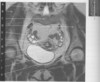
5
Q

figure B 30 arrow A

A

abdominal aorta

6
Q

figure B 30 arrow B

A

splenic vein

7
Q

figure B 30 arrow C

A

portal vein

8
Q

figure B 30 arrow D

A

renal vein

9
Q

figure B 30 arrow E

A

superior mesenteric vein

10
Q

figure B 30 arrow F

A

inferior vena cava

11
Q

figure B 30 arrow G

A

iliac artery

12
Q

vascular imaging of the (venous) abdominal vasculature figure B 30 is typically acquired with

A

ceMRA delayed

13
Q

figure B 31 illustrates vascular imaging of the abdominal vasculature and runoff, this acquisition is acquired with dynamic contrast enhancement and

A

table stepping from the abdomen down to the legs

14
Q

figure B 31 arrow A

A

abdominal aorta

15
Q

figure B 31 arrow B

A

abdominal aortic aneurysm

16
Q

figure B 31 arrow C

A

common iliac artery

17
Q

figure B 31 arrow D

A

femoral artery

18
Q

figure B 31 arrow E

A

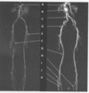
common femoral artery

19
Q

figure B 31 arrow F

A

vascular occlusion

20
Q

figure B 31 arrow G

A

popliteal artery

21
Q

figure B 31 arrow H

A

posterior tibialis artery

22
Q

figure B 31 arrow I

A

anterior tibialis artery

23
Q

figure B 31 arrow J

A

posterior tibialis artery

24
Q

figure B 31 arrow K

A

peroneus brevus artery

25
Q

figure B 32 arrow A

A

subcutaneous fat

26
Q

figure B 32 arrow B

A

rectus abdominus muscle

27
Q

figure B 32 arrow C

A

sacrum

28
Q

figure B 32 arrow D

A

bowel

29
figure B 32 arrow E
myometrium
30
figure B 32 arrow F
junctional zone
31
figure B 32 arrow H
endometrium
32
figure B 32 arrow I
bladder
33
figure B 32 arrow J
cervix
34
figure B 32 arrow K
rectum
35
figure B 33 arrow A
spinal canal
36
figure B 33 arrow B
gluteal muscles
37
figure B 33 arrow C
sacrum
38
figure B 33 arrow D
lilium
39
figure B 33 arrow E
ovaries
40
figure B 33 arrow F
fallopian tubes
41
figure B 33 arrow G
junctional zone
42
figure B 33 arrow H
endometrium
43
figure B 33 arrow I
myometrium
44
figure B 33 arrow J
bladder
45
figure B 33 arrow K
femoral head
153
figure B 34 arrow A
subcutaneous fat
154
figure B 34 arrow B
rectus abdominus muscles
155
figure B 34 arrow C
myometrium
156
figure B 34 arrow D
junctional zone
157
figure B 34 arrow E
endometrium
158
figure B 34 arrow F
roof of the acetabulum
159
figure B 34 arrow G
cervix
160
figure B 34 arrow H
rectum
161
for pelvic imaing the best view for the evaluatoin of the uterus is the
sagittal
162
for femal pelvic imaging the best view for the evaluatoin of the ovaries is the
coronal
163
figure B 35 arrow A
symphysis pubis
164
figure B 35 arrow B
pubic bone
165
figure B 35 arrow C
central gland of the prostate
166
figure B 35 arrow D
peripheral zone of the prostate (with cancer)
167
figure B 35 arrow E
peripheral zone of the prostate (without cancer)
168
figure B 35 arrow F
neurovascular bundle
169
figure B 35 arrow G
obturator muscle
170
figure B 35 arrow H
rectum
171
figure B 36 arrow A
bladder
172
figure B 36 arrow B
seminal vesicles
173
figure B 36 arrow C
vas deferans
174
figure B 36 arrow D
central gland of the prostate
175
figure B 36 arrow E
base of the prostate
176
figure B 36 arrow F
obturator internaus muscle
177
figure B 36 arrow G
apex of the prostate
178
figure B 36 arrow H
prostate urethra
179
figure B 37 arrow A
bowel gas
180
figure B 37 arrow B
seminal vesicles
181
figure B 37 arrow C
rectum
182
figure B 37 arrow D
bladder
183
figure B 37 arrow E
base of the prostate
184
figure B 37 arrow F
peripheral zone of the prostate
185
figure B 37 arrow G
apex of the prostate
186
figure B 37 arrow H
symphysis pubis