44. Endocrine Glands Flashcards
What do endocrine glands do, and how?
Where is the pituitary gland located?
What does the pituitary produce?
How are the anterior and posterior pituitary glands different?
Maintain homeostasis (regulate growth, metabolism etc.) Network of glands, ductless (secrete hormones directly into blood), endocrine organs in cranium, neck, thorax, abdomen and pelvis. NB: exocrine glands have ducts
In sella turcica, middle of sphenoid bone. Hangs of hypothalamus. Optic chiasm in front. [pic]
GOAT FLAP: GH, Oxytocin, ADH, TSH, FSH, LH, ACTH, PRL
Anterior: adenohypophysis, glandular part. Posterior: neurohypophysis, made of neuronal tissue, axons go down infundibulum to posterior PG. (ADH and oxytocin)

What are the 2 nuclei in the posterior pituitary gland, and what do they produce?
How do their hormones enter the blood?
Describe the anterior pituitary blood supply.
Supraoptic (ADH), paraventricular (oxytocin)
Axons transport ADH/oxytocin down and directly into capillary bed.
Supply from internal carotid -> super hypophyseal artery -> hypothalamus -> primary capillary plexus. Hypothalamus secretes hormones into primary capillary plexus -> drains via hypophyseal portal vein to secondary plexus to stimulate anterior PG to secrete e.g. TSH in to efferent hypophyseal veins -> cavernous sinus -> circulation -> thyroid gland -> release T3 and T4

How can a pituitary tumour cause vision problems?
How does the internal carotid artery run through the cavernous sinus?
Pituitary gland under optic chiasm so if tumour -> compresses optic chiasm -> bitemporal hemianopia (tunnel vision)
Runs vertically up (cervical part), enters petrous part of skull base, runs obliquely, enters cavernous sinus and does 180o turn -> into cerebral part of carotid

Label A-F in the cavernous sinus

A) internal carotid
B) CN III (oculomotor)
C) CN IV (trochlear)
D) CNV (trigeminal) - V1 (opthalmic)
E) CNV (trigeminal) - V2 (maxillary)
F) CN VI - (abducens)
Pituitary tumour compresses cranial nerves first
What would be the suspected diagnosis for this patient?

Pituitary tumour so excessive ACTH produced -> cortisol from adrenal glands = Cushing’s like symptoms. Tumour compressing optic chiasm and CNIII, IV and VI = ophthalmoplegia
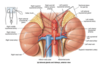
Describe the different parts of the adrenal glands including what they produce.
Where are the adrenal glands located? How are they shaped?
Adrenal cortex: zona glomerulosa (mineralocorticoids - aldosterone), zona fasiculata (glucocorticoids - cortisol), zona reticularis (androgens - testosterone). Adrenal medulla: adrenaline, NA
Between superomedial aspects of kidneys and crura of diaphragm, in perirenal fat. R = pyramidal shaped (in contact with liver and IVC). L = crescent shaped (spleen, stomach and pancreas)

Describe the blood supply to the adrenal glands.
What is the venous supply?
3 differenent BV supply AG: from above (superior suprarenal arteries from inferior phrenic), middle (middle suprarenal artery from abdominal aorta) and bottom (inferior suprarenal artery from renal artery).
Large suprarenal vein (R from IVC, L from L renal vein)

Parathyroid glands -> hypocalcaemia/can’t regulate Ca levels if damaged/removed -> tetany
Recurrant laryngeal b/c runs right behind it and quite small. If damage 1 = hoarse voice, if 2 = dysphonia.
Thyroid ima artery b/c lots of people have it in the midline so need to be careful when making incisions
Label the following structures anterior to the thyroid gland.

A: hyoid
B: thyroid
C: cricoid
D: thyrohyoid
E: sternothyroid
F: omohyoid (attached to scapula - omo = shoulder)
G: sternohyoid
Also suprahyoid = contract and pull up hyoid, larynx follows. Infrahyoid = ovelies the 4 muscles in the picture
What spinal levels does the thyroid gland lie between?
Describe the structure of the thyroid.
C5 - T1
L and R lobe connected by a thin isthmus. V vascular. Pyramidal lobe = remnant of how thyroid developed. Isthmus on top of cricoid cartilage.

Describe the blood supply to the thyroid.
Common carotid either side -> bifurcates to external (supplies face and neck) and internal (supplies brain only) carotid arteries. Superior thyroid artery branches off from external carotid. Inferior thyroid artery branches from thyrocervical trunk. Thyroid ima artery can come from any branch of the arch of the aorta = variable.

Describe the thyroid gland’s venous supply.
What is the parathyroid gland? What is its blood supply?
What does PTH do?
Inferior jugular vein runs along it, joins subclavian. Venous drainage from thyroid goes into these major veins. Inferior thyroid vein drains to brachiocephalic vein. Superior and middle thyroid vein drain to internal jugular vein.
4 (not always) little parathyroid glands embedded in posterior thyroid gland. Superior thyroid arteries supply superior parathyroid and inferior thyroid arteries supply inferior parathyroid gland.
Secreted by parathyroid gland, regulates blood Ca (increases)


Gall stone blocking ampulla of Vata so bile backs up into pancreas -> pancreatitis
What are the 2 jobs of the pancreas?
Describe the pancreas.
Where does the pancreatic blood supply come from?
Exocrine: secretion of powerful digestive enzymes into small intestine
Endocrine: releases insulin and glucagon into blood to determine how body uses food for energy
Retroperitoneal organ, anterior to abdominal aorta, tail in contact with spleen. Head, neck, body, tail, ulcinate process.
Coeliac trunk and superior mesenteric artery. Superior meseteric artery and vein, behind neck but in front of ulcinate process.

Label the blood supply to the pancreas.

A) common hepatic
B) gastroduodenal
C) superior pancreatoduodenal (supplies mostly head)
D) inferior pancreatoduodenal (first banch off superior mesenteric, anastamoses with superior pancreatoduodenal)
E) splenic artery (gives off F, supplies most of body and tail)
F) transverse pancreatic
Label A-G

A) cystic duct
B) L and R hepatic ducts
C) common hepatic duct
D) common bile duct (runs down in free border of lesser omentum)
E) hepatopancreatic ampulla
F) minor duodenal papilla
G) major duodenal papilla (drains tail, body and head of pancreas)
Accessory duct drains ulcinate process
What happens if the gall bladder concentrates bile too much?
Gallstones - only a problem when start to move into duct system -> blockage -> pain and damage. Commonly lodges in ampulla of Vata, back up of bile in biliary tree (pain whenever eay fatty foods). Bile can also back up main pancreatic duct -> acute pancreatitis
