(16.1) Pulmonary Pathology II (Singh) Flashcards
What is a restrictive lung disease?
aka interstitial lung dz, characterized by volume restriction (stiff lungs) = cannot fill the lung
FEV1/FVC ratio is normal or increased (both are reduced BUT the ratio is not necessarily reduced)
(FEV=force expiratory volume, FVC=forced vital capacity)
What is the pathogenesis of Idipoathic pulmonary fibrosis?
IPF damages pulmonary tissue with waves of inflammatory injury leading to fibrosis
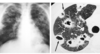
What would a CXR look like for a idiopathic pulmonary fibrosis pt?
Basilar infiltrates
“Honeycomb lung”

What do the lungs sound like on auscultation for idiopathic pulmonary fibrosis?
Crackles on exam
“Velcro-like”
What are the contributing factors to idiopathic pulmonary fibrosis?
Enviornmental factors (SMOKING)
Genetic factors
Increasing age
What is unique about the histology of idiopathic pulmonary fibrosis?
Very different patterns due to the “wave like” nature of the disease
Some patches are normal, some have inflammation, others have fibroblast foci and some have peripheral honeycombing

What do pathologists call idiopathic pulmonary fibrosis when found on pulmonary biopsy?
Usual Interstitial Pneumonia (UIP)

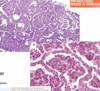
What do these images represent?

Honeycomb fibrosis

What is the prognosis for patients with idiopathic pulmonary fibrosis?
NOT GOOD
Most patients die from respiratory disease 3-5 years after diagnosis (either from respiratory failure or cor pulmonale)
Only truly effective treatment = lung transplant
What are some experimental medications being used to treat idiopathic pulmonary fibrosis?
These meds are used to arrest fibrosis
- Tyrosine kinase inhibitors*
- TGF-Beta inhibitors*
What is non-specific interstitial pneumonia (NSIP)?
VERY SIMILAR TO UIP
Idiopathic
Has UNIQUE HISTOLOGY = uniform infiltrates and fibrosis
Has better prognosis than UIP
What is this lung disorder?

Non-specific interstitial pneumonia (NSIP)

What does cryptogenic organizing pneumonia (COP) looks like histologically?
Looks like cotton candy
The “cotton candy” is fibroblast foci (Masson bodies) = organizing plugs of connective tissue

Cryptogenic organizing pneumonia (COP)
Prognosis?

Very good!
Patient tend to have full recovery with oral steroids since the fibroblast foci are early fibrosis that are so poorly established
How do you diagnose cryptogenic organizing pneumonia (COP)?
Diagnosis of exclusion
-Not an infection, drug- or toxin-induced, or related to connective tissue disorders
What is an important consideration for patients presenting with possible pulmonary fibrosis (IFP, NSIP, COP)?
This may be secondary to their autoimmune/connective tissue disease such as RA, Scleroderma or SLE
important to treat the underlying disorder that is causing the fibrosis
What is this?

Granulomatous inflammation

What is sarcoidosis?
Systemic disease manifesting non-caseating (non-necrotizing) granulomata
What is the clinical presentation of sarcoidosis?
Incidental abnormal radiograph
or
Dyspnea
What are some of the hallmark granuloma inclusions of sarcoidosis?
Granuloma inclusions:
Asteroid body (A)
Schaumann bodies (B-D)

What is the demographic of sarcoidosis?
<40 years of age
African americans
Commonly involve LUNGS
Elevated ACE levels
Sarcoidosis
Do the stages occur in order?
What are the common causes of death in sarcoidosis?
No
from pulmonary, cardiac or neurologic involvement
What is this?

Hypersensitivity Pneumonitis
Granuloma isn’t well defined becuase it is mixed with inflammatory cells as a reaction to inhaled substance
What is hypersensitivity pneumonitis?
Immune reaction to inhaled organic antigens (bird poop, hay, Mycobacterium avium complex)
What are three examples of specific causes of hypersensitivity pneumonitis?
Pigeon-breeder’s lung — protein from bird feces
Farmer’s lung — Actinomycetic spores in hay
Hot tub lung — Reaction to mycobacterium avium complex (MAC)
What is one of the most imporant aspects of your clinical interaction w/ your patient that will allow you to diagnose hypersensitivity pneumonitis?
HISTORY
(smoking, vaping, passive exposure to something, medication?)
What is the pathology?

Desquamative Interstitial Pneumonia (DSIP)
*Notice that every alveolar space has clumps of macrophages inside. Also, a MISNOMER, these are NOT squamous cells

What is the demographic of desquamative interstitial pneumonia (DSIP)?
Smokers in 40s/50s
Restrictive lung disease presentation
What is the prognosis for desquamative interstitial pneumonia (DSIP)?
Good prognosis only IF THE PATIENT CAN STOP SMOKING
can also be treated with corticosteroids
What is the pathology?

Respiratory Bronchiolitis-Interstitial Lung Disease (RB-ILD)

What is the demographic for respiratory bonchiolitis-interstitial lung disease?
Smokers
30s-40s
What is the pathology?

Langerhans cell histiocytosis (LCH)

What is the pathogenesis of LCH?
progressive scarring leading to cysts that could rupture and become a pneumothorax
this dz is classically seen in young smokers, can reverse with smoking cessation
What is a major characteristic of langerhans cells histologically?
Langerhans cells ALWAYS stain CD1a positive
What are some dead giveaways that you are looking at a langerhans cell histiocytosis (LCH) biopsy?
Langerhans cells with a TON of eosinophils (red tinges)

What is pulmonary alveolar proteinosis?
autoimmune Impairment of surfactant metabolism due to defect in granulocyte-macrophage colony stimulating factor (GM-CSF) > too much surfactant
What is the histology of pulmonary alveolar proeinosis?
*Frequently confused with pulmonary edema

How do you treat pulmonary alveolar proteinosis?
Treat with SubQ GM-CSF or bronchioalveolar lavage to take out all the extra surfactant
What is this?

Bone marrow embolism

What is this?

Talc embolism
Seen in IV drug users

What is this?

Septic emboli

What is pneumoconiosis?
Reaction by the lungs to inhaled mineral or organic dust
*occupational exposure, air pollution
What factors can worsen pneumoconiosis?
high and repetitive exposure, small particles reaching alveoli, impaired ciliary clearance (eg smoking)
What is coal worker’s pneumoconiosis?
What are the complications?
Black lung due to coal dust
Anthracosis (carbon laden marchophages), coal macules/nodules, progressive massive fibrosis (usually not that severe, but can progress to respiratory failure in some cases)

What is silicosis?
Disease resulting from inhaled silicon dioxide, increases risk of lung cancer
*mining/quarry work/concrete repair/demolition
Describe the histologic findings for silicosis
Dense collagenous nodules

Describe the radiographic findings of silicosis
Eggshell calcifications
(calcified hilar lymph nodes)

What populations are at risk for asbestosis exposure?
What symptoms can be seen?
Insulation workers, Shipyard workers, Paper mill workers, Oil or chemical refinery workers
Pleural (plaques, effusions, mesothelioma) or lung (interstitial fibrosis, carcinoma) manifestations
What are histologic hallmarks of asbestos inhalation?
Asbestos bodies

What cancer is highly associated with asbestos exposure?
Mesothelioma, may present decades later

What is the downstream consequence of a PE?
Pulmonary infarct
wedge shaped lesions that begins as hemorrhagic and becomes fibrotic

If a PE is larger, what is a consequence?
What is seen post mortem in these patients?
Saddle pulmonary embolism - blockage at the pulmoanary trunk leading to acute cor pulmonale and cardiac death
Lines of Zahn - alternating pale (platelet/fibrin) and red (red cells) areas, indicates that embolus occurred while patient is alive (antemortem) and is the cause of death
What can cause these lung nodules/abscesses in IV drug users?

septic embolus coming from a infected heart valves secondary to IV drug use > can form abscesses in the lung parenchyma
suspect endocarditis, FROM JANE symptoms
What is the definition of pulmonary hypertension?
Pulmonary artery pressure (PAP) is greater than 25mmHg
What are the two histological manifestations of pulmonary hyptertension?
Plexiform lesion (left)
Medial hypertrophy (right)

What is the pathology?

Pulmonary hemorrhage syndromes

What is the pathology?

Goodpasture syndrome
Anti-basement antibodies (igG) deposit on the basement membrane of the lung parenchyma + RBCs within the alveolar spaces (hemorrhaging)

What is goodpasture syndrome?
Antibody-mediated disease that damages basement membranes in the lung and kidney
*Targets Collagen IV
What is the demographic of goodpasture syndrome?
YOUNG MEN in their 20s/30s, may present with lung hemorrhaging (red lung) + renal issues
What is the pathology?

GPA : Granulomatosis w/ polyangiitis (huge masses formed by the vasculitis in lung vessels)

__________ is the second most common cause of hospital admissions
Pneumonia
What is the diagnosis?

Right upper lobar pneumonia (consolidated area in the right upper lobe)

What are the 4 stages of lobar pneumonia?
- Congestion (vascular engorgement due to increased blood flow)
- Red hapatization (red cells and inflammation)
- Grey hepatization (inflammation and debris)
- Resolution (fibrosis, macrophage clean-up)

What is the most common cause of community acquired pneumonia?
Who needs to be vaccinated against this organism?
Streptococcus pneumonia (Lancet shaped gram positive diplococci)
infants,>65 yo, respiratory dz, smokers
What are the complications of lobar pneumonia?
Abscess formation that can spread to the pleura (empyema) and systemic circulation (bacteremia)
*lobar pneumonia is usually bacterial etiology

What is the most common cause of CAP in children?
in IV drug users?
in alcoholics?
in CF patients?
H. influenzae
S. aureus (also abscess formation)
K. pneumoniae (currant jelly sputum)
P. aureginosa (copper rust/greenish tinge on sputum and tissue)
What is “atypical” (“walking”) pneumonia?
Slower onset than typical pneumonia
Systemic symptoms predominate
Patchy infiltrates on CXR
Young adults/teens/older children
What is the major bacterium that causes “atypical walking pneumonia”?
Mycoplasma pneumoniae
What is so special about mycoplasma pneumoniae?
Smallest free-living, self-replicating microorganisms
NO CELL WALL

Where would you find legionella pneumophila?
Grows in warm freshwater (gram neg bacillus)
- Air conditioning units
- Misters
- Hot tubs
What are the three major causes of community acquired viral pneumonia?
Influenza (H1N1)
SARS
Respiratory Syncytial Virus
How are influenza viruses classified?
Possession of these proteins:
Hemagglutinin (anchors to the cell)
Neuraminidase (cleaves the hemagluttinin achor to release the virions to infect other cells)

What is antigenic drift?
causes epidemics
MINOR changes to proteins on the virus, allowing increased spread
Similar enough to the orignial virus to allow for some immunity in many individuals
What is antigenic shift?
Pandemics
Genomic alterations with MAJOR resulting changes to protein structure
Naive immunity for almost all people
***Remember = Shift is close to “shit”. Antigenic shit. Antigenic shift is WAY worse than drift.
What is the pathogenesis of community acquired viral pneumonia?
What is a complication of viral pneumonia?
Virus enters the lung cell > cytokine storm > tissue damage/edema > fluid buildup in the alveoli + hyaline membranes (ARDS)
Damaged lungs vulnerable to a bacterial pneumonia which can ultimately cause death
What is COVID-19?
What is an interesting complication associated with this?
+ sense ssRNA, spread via respiratory droplets > induces cytokine storm leading to ARDS
systemic coagulopathy (arterial/venous thrombus formation + megakaryocytes + elevated D-dimer)
What are the bacterial associations you should make with neonates?
Group B strep
Gram negative bacilli
Listeria
*acquired from the birthing canal
What viral and bacterial associations should you make with children >1month old?
Viral = RESPIRATORY SYNCYTIAL VIRUS, parainfluenza virus, Influenza A&B, Adenovirus, Rhinovirus
Bacterial = S.pneumoniae, H. influenzae, M. catarrhalis, S.aureus
What is the pathology?

Respiratory Syncytial Virus (RSV)

What are the symptoms of respiratory syncytial virus?
Rhinorrhea/cough
Wheezing
Dyspnea
Tachypnea
Cyanosis
Histologically, what is the difference b/w bacterial and viral pneumonia?
Bacterial usually stays in alveolar spaces
Viral stays in the interstitum

What are the differences b/w bacterial and viral pneumonia in terms of clinical presentation?
Bacterial: abrupt, high fever, crackles, lobar
Viral: gradual, epidemics, wheezing, diffuse ifiltrates
What are lung abscesses?
Complication of pneumonia (usually due to S. aureus, K pneumoniae)
Also seen in aspiration pneumonia (alcoholics, elderly stroke pts and anaerobic bacterial pneumonia)
What is the pathogenesis of tuberculosis?
Caseating granuloma in lung + hilar nodes (ghon complex)
Usually heals with calcification and fibrosis = latent TB
If patient becomes immunosuppressed: latent TB > secondary TB > could lead to miliary TB
What pathology is HIGHLY associated with TB?
Caseating granulomata

What is the pathology?

Histoplasma capsulatum

Where is histoplasma capsulatum endemic?
Midwest and caribbean
What is the typical clinical presentation of histoplasma?
What is seen on histology?
subclinical/mild infection, can appear as calcifications or coin lesions in CXR with granulomas, but can be aggressive in immunocompromised
Pumpkin seed morphology

What is this infection?
How will this present?

Blastomyces dermatitides - broad based budding
granulomatous pulmonary infection, can also spread to the skin
What is this infection?
How will this present?

Coccidiodes immitis
granulomatous infection with eosinophils, often subclinical unless immunocompromised

Where is coccidiodes immitis endemic?
Southwestern US and Mexico
What is this?

Pneumocystis jiroveci (cup shaped yeast)

What is pneumocystis jiroveci associated with?
AIDS (only manifests in immunocompromised patients)
What is this?

Mycobacterium avium complex (MAC) - acid fast staining
Who typically gets mycobacterium avium complex?
Immunocompromised or elderly
Why is an important consideration with transplant patients presenting with systemic symptoms?
Patient could have rejection (if you get too little rejection meds), OR have an opportunistic infection (too much rejection meds)
Determining which is occuring via biopsy will determine the course of action
Tx for acute rejection = increase immunosuppression
Tx for opportunistic infection = target the organism
