1 - Chest Imaging Flashcards
What is the ideal orientation for chest x ray?
P-A
Heart closer to detector
Better lung image
Structure

Right Para-Tracheal Stripe
Thickening may indicate mass or enlarged lymph node
Left side not well defined dur to aortic arch

Window
How is heart sized assessed on radiographs?
What is the only orientation you’d use?
Ratio of widest diameters of heart to widest internal diameter of thoracic cage
Normal is < 50%
- - -
P-A


Pneumoperitoneum
Free Air at arrow
What condition is happening at the arrows?

The diaphragm is flattened, note the flattened costophrenic angles
Can be caused by emphysema
What’s going on at the arrow you jackass?

Effusion, costophrenic recess
Look for blunted angles
What is going on in this image?

- Heart and mediastinum shifted left
- Absent lung markings at upper arrows
- Collapsed right lung
= Tension Pneumothorax

Upper - Retrosternal Space
Lucent area b/t the sternum and ascending aorta/hear
Lower - Retrocardiac Space
Posterior to posterior heart border; can decrease with enlargement of posterior of the heart

Which of these is bad?

Left - Anterior Mediastinal Adenopathy
Right is just a f*cking arm.
What view do you need to assess the hila?
Lateral

What’s going on here buster?

Lower left lobe pneumonia is superimposed on the lower spine at the white arrow
Called “Spine Sign”

Fluid blunting the posterior costophrenic sulcus (white arrow)
Pleural effusion is on the right
On a lung window CT, what color will the lungs be?
What would indicate a mass in the lungs?
Black
large areas of white may indicate lesion or mass
Five Vessel Level

Trachea - T - Black from air, circular
Esophagus - not labeled - Posterior to trachea, usually collapsed
Great vessels:
R Brachiocephalic V - RBV
L Brachiocephalic V - LBV
Brachiocephalic Trunk - BT
Common Carotid - C
Left Subclavian - S

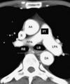
Aortic Arch Level

Aortic Arch - aa
Superior Vena Cava - S
Azygous Vein - A
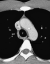
Aortopulmonary Window Level

Ascending Aorta - AA
Descending Aorta - DA
Superior Vena Cava - S
uppermost aspect of left pulmonary - P
Arrow = Aortopulmonary Window

Main Pulmonary Artery Level

Superior Vena Cava - S
Ascending Aorta - AA
Right and Left Pulmonary Arteries - RPA, LPA
Pulmonary Trunk - PT
Right and Left Main Bronchi - RB, LB (black from air, circular)
Descending Aorta - DA
High Cardiac Level

Left Atrium - LA (posterior heart, note pulmonary veins entering)
Right Atrium - RA (right border)
Ascending Aorta - AA
Right Ventricle - RV (anterior border)
Left Ventricle - LV
Descending Aorta - DA

Low Cardiac Level

Right Atrium - RA
Left Atrium - LA
Right Ventricle - RV
Left Ventricle - LV
Pericardium - White arrow, usually 2mm thick
Interventricular Septum - IVS black arrow
Descending Aorta - DA

