Week 8: Urine Concentrating and Diluting Mechanisms Flashcards
(37 cards)
What osmolality (hyper,iso,hypo osmotic) is the filtrate in the PCT?
isosmotic

What osmolality (hyper,iso,hypo osmotic) is the filtrate in the thin part of the descending loop?
Water is reabsorbed and therefore it is hyperosmotic (as water goes out without salt reabsorption)

What osmolality (hyper,iso,hypo osmotic) is the filtrate in the Loop of Henle?
Water is reabsorbed and therefore it is hyperosmotic (as water goes out without salt reabsorption)

What osmolality (hyper,iso,hypo osmotic) is the filtrate in the thick ascending limb?
Hypoosmotic
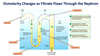
What osmolality (hyper,iso,hypo osmotic) is the filtrate in the DCT?
Hypoosmotic

What osmolality (hyper,iso,hypo osmotic) is the filtrate in the Collecting duct?
variable - changes depending on ADH. If there is increase in ADH the solute will become hyperosmotic as water is being reabsorbed. If no ADH then urine will be dilute as water is remaining in urine.

Urine is considered concentrated near osmolality _____ , and dilute when closer to ______.
1200mOsm, 50 mOsm
What cell type is acted upon by ADH and is responsible for water reabsorption in the collecting duct?
Principle Cells
Describe the stimulation mechanism for the production of ADH pathway (there are 3 and then the site it acts on.

Describe the machanism of how ADH increases water reabsorption in the dollecting duct (on a cellular level) (HINT: there’s 5 steps)
- Vasopressin binds with its receptor site on the basolateral membrane of a principle cell in the late distal or collecting tubule
- The binding activates cAMP within the cell as a second messenger
- cAMP then promotes the insertion of Aquaporin 2 into the cells apical membrane (which is usually impermeable to water)
- water then freely enters into the tubular cell from the lumen through these aquaporin 2 channels
- Water then exits the cell into the blood through Aquaporin 3 and 4 (which are permanently positioned at this basolateral border, and not influenced by ADH)
- Water is thus transported into the blood and is successfully reabsorbed

What happens to the aquaporin 2 channels when stimulated by ADH and when there is no ADH?
- These aquaporin 2 channels are produced and then exocytosed into the membrane (while stimulated by ADH) to be incorporated
- Once the ADH stimulation goes, these aquaporins are endocytosed back into the cell no longer allowing the passage of water

Within the kidney, what are teh 2 types of water reabsoprtion?
- Obligatory Water Reabsorption
- Is water movement that cannot be prevented
- This is due to the movement of solutes which alters osmolarity forcing osmotic water reabsorption
- This usually recovers around 85% of the filtrate produced - Facultative Water Reabsorption
- This is water reabsorption that is assisted by some other factor, in this case hormones
- This type controls volumes of water reabsorbed along the DCT and Collecting Ducts through the action of ADH
- This usually reabsorbs around 15% of filtrate
How is “free water” made in the kidneys (water that is free of solute) and how does positive and negative free water clearance occur?
- Within the thick ascending limb and distal convoluted tubule, the region known as the diluting segment, the filtrate is diluted as they are impermeable to water, and only allow the reabsorption of solutes
- This generates an amount of free water (water that is free of solutes)
- If this free water is excreted (no ADH), the urine will be hypoosmotic (dilute) which means free water clearance is positive
- If this free water is reabsorbed in the late distal tubule and collecting duct, the urine will be hyperosmotic meaning the free water clearance will be negative

Explain the mechanism of excretion of dilute urine throughout the tubular system
- The filtrate that enters the tubular system will continue down the Loop of Henle, and water will be absorbed
- As it comes back up into the diluting segment (thick ascending limb and distal convoluted tubule), water is impermeable and so only solutes will leave
- This will dilute the fluid
- If this dilute fluid enters into the late distal tubule and collecting tubule in the absence of ADH, solutes will continue to be reabsorbed, further diluting the filtrate
- As no water is reabsorbed, their will be a large volume of urine with a very low osmolality
- Basically, without ADH, water is not reabsorbed in the distal tubule, thus all fluid reaching this DCT is lost in urine, producing large amounts of dilute urine

Explain the mechanism of concentrating urine in the tubular system (HINT: through ADH)
- Tubular fluid enters the late distal tubule with an osmolarity of 100mOsmm
- In the presence of ADH, the principal cells of late distal tubule and cortical collecting tubule are permeable to water, water thus moves into the interstitium of the cortex, and is carried away by the peritubular capillaries of the vasa recta
- The filtrate continues, and more water reabsorption occurs in the medullary collecting ducts, at which point the osmolality of the fluid is 1200mOsm
- This process is increased by increased urea and NaCl in medulla (as it increases medulla interstitium osmolarity)

What is obligatory urine volume and what is the minimum amount that must be excreted each day (in mL and mOsm)
a minimal volume of urine that must be excreted to rid the body of waste products. This minimum amount is 500mL/day or must excrete about 600mOsm of solute each day.
Obligatory urine volume is increased in ___ disease, as a result of _______
kidney, impaired urine concentrating ability
What is the osmolarity in the medulla
1200mOsm - hyperosmolarity
What are the 2 operations which increase osmolarity in the medulla and where are they located?
- Counter current exchanger - loop of henle
- Counter current multiplier - vasa recta
occurs in the loop of henle with the vasa recta
What are the 4 factors contributing to medullary hyperosmolarity?
- Sodium-Potassium-2-chloride symporters in the thick ascending limb
- Through this transporter salt is reabsorbed adding sodium chloride to the medullary interstitium - Principal cells in Collecting ducts increase sodium reabsorption into the medullary interstitium through the action of aldosterone (ENaCs)
- This active transportation further adds salt to the medullary interstitium - Facilitated diffusion by ADH of large amounts of UREA in the medullary collecting ducts
- Any difussion of small amounts of water from the medullary tubules which leaks into the medullary interstitium is taken by the vasa recta.
Simply put, salt and UREA create the hyperosmolality of the medullary interstitium which is essential for the concentration mechanism
Explain the countercurrent multiplier
- More salt is continually added by the Proximal Convoluted Tubule (by filtration)
- The higher the osmolarity of the ECF, the more water that will follow, leaving the descending limb by osmosis
- The more water that leaves the descending limb, the saltier the fluid is that remains in the tubule
- The saltier the fluid in the ascending limb, the more salt the tubule will pump out into the ECF
- The more salt that is pumped out of the ascending limb, the saltier the ECF is in the renal medulla (go to step 2 and repeat)
- Basically, the long nephron loop creates a gradient, and a positive feedback cycle uses the flow of fluid to multiply and constantly replenish the medullary interstitial fluid concentrations

Explain the countercurrent exchanger
- The blood osmolality of the descending loop is around 300mOsm and so as it comes down, it enters into the hyperosmolality region which has very high concentration of sodium chloride and UREA
- As it is a blood vessel, this salt crosses over into the blood vessel (from the medulla) and water goes out as it is in a state of hyperosmolality outside (meaning water wants to dilute it)
- As the blood then moves down the vasa recta It takes all of this sodium chloride until the concentration is equal to that of the interstitial medullary fluid (it is hyperosmolar), there is equilibrium (the salt wants to move from a high concentration to a low concentration)
- As blood flows out, it flows upwards into the regions of the reduced osmolality, the 1200mOsm in the blood is greater then the osmolality of the interstitium
- This causes the sodium chloride within the vessel to move out into the interstitium down its concentration gradient, whilst drawing water back in from the interstitium to dilute the osmolality of the vessel
- Thus, as blood flows up and out, it leaves behind the sodium chloride and takes away the water (takes a little bit, that is ends up with an osmolality of around 325mOsm)
- The net effect is the maintenance of medullary interstitium hyperosmolality

Explain the recirculation of UREA
- UREA is initially passively reabsorbed within the PCT
- ADH increases UREA reabsorption at the Collecting duct (through urea transporter UT-A1 and UT-A3)
- This causes a high concentration of UREA in the medullary interstitium
- This UREA adds to the medullary interstitial osmolality
- It is then secreted into the loop of Henle (via UT-A2, which isn’t hormone dependent), it then goes up and comes back to the collecting duct, and is reabsorbed once more creating a recirculation effect
- This UREA provides 600mOsm to medullary interstitium

Explain the mechanism of what would happen if we were overhydrated


