Week 3-Lumbar puncture, epidural, spinal and caudal Flashcards
What are the indications for a lumbar puncture?
Diagnosis of:
meningitis/ encephalitis
subarachnoid haemorrhage (in patient with normal CT)
measurement of CSF pressure
removal of CSF therapeutically (idiopathic cranial hypertension)
diagnosis of MS/ neurosyphilis
intrathecal injections and drugs
What are the normal parameters of CSF?
normal parameters of CSF:
appearance –> crystal clear, colourless
pressure –> 8-15 mmHg
cell count –> no polymorphs, mononuclear cells only, less than 5/mm3
protein 0.2-0.4 g/L
Glucose 2/3 - 1/2 of blood glucose
IgG –> less than 15% totatl CSF protein
oligoclonal bands –> absent
What are the contraindications to LP?
- Patient refusal
- Raised intracranial pressure –> papilloedema via fundoscopy, bulging optic disc and engorged veins
- Regional skin/ soft tissue infection
- cord compression
- coagulopathy
- congenital malformation
- spinal fixation surgery

How is a patient checked for raised ICP?
What are the complications of LP?
Headache (common)
Bloody (traumatic) tap
Brain or cerebellar herniation
Extradural haematoma (within fat filled space with veins, bleed can track up and compress the spinal cord).
Meningitis
CN VI palsy/ hearing loss
Transient/ persistent paraesthesia/ anaesthesia

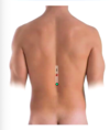
Label image
What should you be careful of during a LP?

1) Vertebral body
2) vertebral disc –> nucleus pulposus, annulus fibrosis
3) pedicle (directly posterior to vertebral body)
4) spinous process
5) Transverse process
Ligaments (from outside to in):
1) supraspinous ligament
2) interspinous ligament
3) ligamentum flavum (elastic, yellow coloured)
Need to be careful not to puncture too far through the ligamentum flavum and not to hit the intervertebral disc –> could increase risk of disc herniation and pain for the pt.
Where does the spinal cord terminate?
What level is lumbar puncture done at?
Spinal cord terminates at L1/L2
Lumbar puncture is done normally at L3/L4 (although can be done at L4/L5, L5/S1 as there is still CSF) to avoid the end of the spinal cord (conus medullaris). The cauda equina nerves tend to move out the way of the needle.
Where does the CSF filled sub arachnoid space terminate in most people?
What is the range of this?
What can the termination of subarachnoid space be landmarked by?
CSF filled subcarachnoid space terminates in 90% of adults from the lower part of S1-S2 level
(ranges from L5-S1 to S4).
Termination of the subarachnoid space can be landmarked using the PSIS and sacral spinous processes.

Label the image -> what are the meningeal layers and spaces inbetween?
what are the contents of these spaces?

Spinal cord is immediately surrounded by the pia mater
Outside the pia mater is the subarachnoid space (filled with CSF and connective tissue trabeculae)
Around the subarachnoid space is the arachnoid mater
outside the arachnoid mater is the subdural space with traversing cerebral bridging veins, draining the neural tissue into the dural sinuses (potential space, can be opened by separation of arachnoid mater from the dura mater by result of trauma, pathologic process, or the absence of cerebrospinal fluid as seen in a cadaver)
outside the subdural space is the dura mater
outside the dura mater is the epidural space filled with adipose tissue and the vertebral venous plexus

What structures do you need to be careful of during LP?
Spinal nerves leave the spinal cord laterally, you need to be careful not to impact a spinal nerve as you enter the needle.

What are the vertebral levels related to these two points?


What does the cauda equina consist of?
What space does it occupy?
Cauda equina consists of nerves and nerve rootlets from:
L2-L5
S1-S5
coccygeal nerve
Cauda equina occupies the lumbar cistern, the subarachnoid space inferior to the conus medullaris
Is this slice above or below L1?
Would a LP be done here?
How can you tell? (label as many features as poss)

This slice is above the level of L1 as the spinal cord is still present.
Immediately surrounding the spinal cord are the meningeal layers, the red line consisting of both the arachnoid and dura mater.
Outside the dura mater is the epidural space filled with fat and venous plexus (the dark fragments)
Either side of the spinal cord you can see two spinal nerves exiting
In front of the spinal cord you can see the large vertebral body and a slice through the vertebral disc (with annulus fibrosus and nucleus pulpolsus).
Posteriorly you can see the spinous process and transverse processes of the lumbar vertebrae.

What does 1) the white zone represent?
What does 2) the red zone show?
Zone 1 –> white box shows the zone of spinal cord termination which can extend from the middle 1/3rd of T11 to the middle 1/3rd of L3
Zone 2 –> red box shows the most common level of spinal cord termination –> at the middle 1/3rd of the L1 vertebral body which corresponds with the interspinous space between T12 and L1 spinoous process (at spinous processes are angled downwards).

What are zones 3 and 4?
Why might doing a spinal tap in zone 3 potentially cause a problem?

Zone 4 –> is the highest point of the iliac crest and the supracristal plane; it intersects the vertebral column from L4 to the the L4/L5 intervertebral disc (in majority of pts).
Find L4 to do your LP, however the zone that palpation directs you to may vary between clinicians, plus anatomical variation between patients leads to zone 3:
Zone of supracristal plane intersection with the vertebral column
Can range from L2-L3 to the L4/L5 interspinous space –> this could potentially cause a problem in a patient with a low terminating cord (at L3). Therefore advice is to always go a vertebral level lower.

What is the yellow part of the spinal cord shown? What spinal nerves arise here? What vertebral level?

The yellow part of the spinal cord = lumbar part
Lumbar spinal nerves arise here, L1-L5, origin actually at T12 vertebral level.
What nerves are the orange part of the spinal cord shown?
What vertebral level is this part of the spinal cord located at?

The orange part of the spinal cord is the sacral part, sacral nerves can be followed exiting sacral foramina.
The sacral portion of the spinal cord can actually be located at L1/L2 (S1-S5 spinal nerves originate here).
What is the range of vertebral levels at which is is acceptable to do LP in and adult?
How does this change in a child?
In adults LP is performed via the L3-L4 to L5-S1 interspinous gap
Preference at L4/L5 vertebral level.
In children and neonates –> lumbar puncture is performed at the L4/L5 of L5-S1 interspinous gap.
This is because the infanct spinal cord terminates at the L3 level

Where is a needle inserted during an LP?
What way should the needle be angled?
Without this angulation what could be hit?
The needle is inserted between spinous processes and is angled anterosuperiorly (15 degrees cephalad orientation, aim towards umbilicus).
Without antero superior angulation, the needle will hit the bone, usually the lamina.

What position should the patient be in during the LP and why?
During LP patient is either on their side or sat on the bed hunched forwards to flex the vertebral column, legs to chest.
This increases the space inbetween the adjacent spinous processes making it easier to insert the needle.
If done on neonates or unconcious patients another person is required to help position them with their vertebral column flexed.

Why/ when might a patient’s O2 sats need monitoring during a LP?
During an LP when a patient is hunched over/ lying down you may reduce tidal volume.
This shouldn’t be problematic in normal patients but in obese patients/ elderly/ patients with comorbidities they can become hypoxic.
What layers are passed through during an LP?
1) Skin and subcutaneous tissue
2) Supraspinous ligament (will feel resistance)
3) interspinous ligament
4) Ligamentum flavum –> feel a loss of resistance/ “give” when passed through

what does the supraspinous ligament become in the cervical region?
Why is this structure surgically useful?
supraspinous ligament becomes the ligamentum nuchae in the cervical region.
Ligamentum nuchae is avascular and aneural, useful for cervical procedures.
What must the needle penetrate (apart from skin/ subcut fat) to reach the subarachnoid space?
When is a loss of resistance/ pop felt?
What space has been entered?
When might another loss of resistance be felt?
What space has been entered?
The needle must penetrate ligamentum flavum, the dura and arachnoid mater to reach the CSF.
Distinct loss of resistance may be felt when needle penetrates ligamentum flavum and enters the epidural space.
Another loss of resistance may be felt as the needle penetrates the dura and arachnoid mater. Now entered the subarachnoid space.


