Week 2-Neck anatomy 1 Flashcards
What three regions does the neck connect?
What is it a collection of?
The neck connects the head upper thorax and upper limb
The neck is a collection of spaces and compartments separated by fascia.
Describe the superior boundary of the neck
Describe the inferior boundary of the neck
Superior boundary of the neck formed by the inferior mandible and base of the skill, along the pericranial line.
Inferior boundary of the neck: Extends down to the manubrium, the clavicle and acromion to the spinous process of C7. (Most prominent process in cervical spine)

What does the neck communicate freely with?
Why is this important?
The neck communicates freely with the thorax and mediastinum.
This is important when considering infection spread from the neck.
What divides the neck into two anatomical regions?
What are these regions called?
What do the muscles do?
Hyoid bone divides the neck into two regions.
Above hyoid = suprahyoid and muscle action here elevates the hyoid
Below hyoid = Infrahyoid, muscle action here depresses the hyoid

Label and decribe the muscle shown
What is the innervation of the muscle?

Muscle shown = Digastric, with anterior and posterior belly attached by central tendon. Central tendon is attached to the hyoid bone by a loop of tissue meaning it can elevate the hyoid.
Innervation: Different nerves innervate different muscle bellies due to embryological development from different pharyngeal arches.
Digastric anterior belly -> CN V3/c
Digastric posterior belly –> CN VII
Label and describe innervation of muscles shown

Large sheet of muscle = Mylohyoid, innervation CN V3/c
Muscle superior to the posterior belly of digastric = Stylohyoid, innervation CNVII
What is the naming convention of the muscles in the neck?
Many neck structures are named according to their attachments from inferior to superior
E.g: Thyrohyoid muscle (from thyroid cartilage to hyoid bone)
Sternothyroid (from sternum to the thryoid cartilage)
Sternohyoid (from sternum to hyoid bone)

Label the grey boxes: What are the muscles?

Omohyoid forms some of the boundaries of the triangles of the neck
Has both a superior belly and inferior belly.
Scalene muscles: anterior, middle and posterior scalenes.
What is the main nerve supply to many of the infrahyoid muscles?
What is the exception?
Main nerve supply to infrahyoid muscles is from the ansa (leather loop straps on sandal) cervicalis (C1-C3).
It is a fine loop of nerves that are part of the cervical plexus. Lies superficial to internal jugular vein in the carotid triangle.
Innervates most infrahyoid muscles: sternothyroid, sternohyoid, omohyoid.
Note thyrohyoid muscle is innervated by C1 spinal nerve via hypoglossal nerve.
What can be damaged during a carotid endartectomy?
What is the consequence?
The ansa cervicalis is often cut during carotid endarterectomy.
(carotid endarterectomy –> removal of atheromatous plaque in common carotid/ internal carotid to reduce risk of stroke and corrects stenosis).
Consequence of cutting ansa cervicalis is relatively minimal, may have dysphagia and dysphonia.
What do posterior neck muscles do?
Why are they quite commonly injured?
Posteriorly located muscles extend or laterally flex the neck, also supports the head against gravity.
As they are under tension most of the time they are quite commonly injured.
Muscular strain/ tears are a cause of posterior neck pain.
Label this structure

- Ligamentum nuchae –> part of supraspinous ligament
- Surgically useful –> provides avascular and aneural plane that can be cut to access the cervical spine
Label the image


Where does the suboccipital triangle sit?
What artery is located here?
What nerves are located here?
Suboccipital triangle sits under the occipital bone in the upper cervical region.
The vertebral artery sits here and is vulnerable to rupture during forced extension of head and neck –> potentially fatal.
C1-C3 nerves here. C1 has no sensory innervation but C2/C3 can.
Entrapment of C2 and C3 dorsal rami can produce a posterior headache / occipital neuralgia.
Occipital neuralgia = consistent and persistent very painful headaches, Tx surgically by releasing muscle entrapment.

What are the two main triangles of the neck?
What are there boundaries?
Neck is divided into 2 main triangles –> anterior and posterior
Anterior triangle boundaries:
Medial border = midline/ medial sagittal plane
Lateral border = Sternocleidomastoid
Superior border = inferior margin of mandible
Posterior triangle boundaries:
Medial = sternocleidomastoid (posterior border)
Lateral= Trapezius
Base = Clavicle (middle 1/3rd)
Apex = mastoid process

What forms the border of the anterior triangle?
What is it further divided into?
The anterior triangle borders are formed by the hyoid bone, the mandible and sternocleidomastoid. It is further divided into subtriangles: The submental triangle and submandibular triangle.

What are the boundaries of the submandibular triangle?
What are the boundaries of the submental triangle?
What is associated with both of these triangles?
Submandibular triangle boundaries are formed by the mandible and the two bellies of the diagastric muscle.
Submental triangle is formed by the hyoid bone, the anterior belly of digastric and the midline. OR can be defined as having two anterior bellies of digastric and the hyoid bone.
Associated with both are lymph nodes: Submandibular lymph nodes in submandibular triangle and submental lymph nodes in submental triangle.

Label the triangles of the neck


Anterior triangle subdivisions: Carotid triangle
What are the boundaries?
Carotid triangle:
Anterior boundary –> Omohyoid (superior belly)
Superior boundary = Digastric (Posterior belly)
Posterior boundary –> Sternocleidomastoid

Which landmarks are used to locate and examine the carotid artery?
What major structures pass through the carotid triangle?
Clinical relevance of carotid triangle?
Lateral to the trachea/ larynx, sits between larynx, trachea and mastoid.
Structures: common carotid, bifurcates around C3/4/ at the upper border of the thyroid cartilage into the internal and external carotid arteries.
Plus internal jugular vein, hypoglossal and vagus nerves.
Clinical relevance:
- many of vessels nerves are superficial and are accessed for surgery
- carotid triangle also contains carotid sinus (dilated portion of internal/ common carotid), contains baroreceptors that detect BP. CN IX feeds this info to the brain and regulates BP.
- In some patients baroreceptors are hypersenstive to stretch, external pressure can cause slowing of HR and decreased BP, underperfusion of the brain and syncope.

Anterior triangle subdivisions: Submandibular triangle
Borders?
What structures are located here?
Borders: Superior border = mandible
Inferior = two bellies of digastric (anterior and posterior)
Structures:
Submandibular lymph nodes, submandibular salivary glands, facial artery and vein also pass through here.

Anterior triangle subdivisions: Submental triangle
Borders?
Structures passing through?
Borders:
- Inferior –> Hyoid bone
- lateral –> Digastric anterior belly
- Medial –> Midline (if halved)
(Base of submental triangle bound by mylohyoid muscle, running from mandible to hyoid bone).
Structures:
- Submental lymph nodes (filter lymph draining from floor of the mouth/ tongue)
*

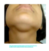
In which region does this mass sit?
What are some differentials?

Submental region mass
Differentials: Submental lymphadenopathy –> due to infection of mandibular teeth, floor of the mouth, gums, lips etc.
Diagnosis: Epidermoid cyst –> due to the fact its movable, squishy and moves with the skin
In which region/s are these neck lumps?
What is the likely cause of the lumps?

Submental, and submandibular triangle masses, plus deep lymph node group : jugulodigastric (where the jular vein is crossed by digastric muscle).
Cause: submental, submandibular and jugulodigastric lymphadenopathy


