Week 2 Overview of Neuropathology (NS Response to Injury) Flashcards
(29 cards)
Developmental
Onset: At birth
Duration: Static
Focality:
Cellular changes: Neuralation or neuronal migration abnormalities
Other Clues: Folate deficiency (valproate)
Trauma
Onset:
Duration: Static
Focality: Yes
Cellular changes: Tissue disruptio, axonal spheroids , petechial hemorhhage (contusion); cell death
Other Clues: Trauma history, suspicious history for pediatric non-accidental trauma
Cerebro-Vasular
Onset: Acute
Duration: Static
Focality: Vascular distribution
Cellular changes: Ischemia and or hemorrhage neurons > neutrophils > macrophages (cavitation)
Other Clues: Risk factors, cardiac arrythmias, atheroscleorisis, hypertension
Toxic metabolic
Onset: subacute
Duration:
Focality: usually global (rarely focal or systemic)
Cellular changes: Alzheimer’s tyoe II astricytes with hepatic encephalopathy
Other Clues: Drugs, vitamin deficiency
Neoplastic
Onset: subacute
Duration: progressive
Focality: usually focal (or regionally infiltrative)
Cellular changes: Atypia, mitoses
Other Clues: Imaging
Infectious
Onset: subacute
Duration: progressive
Focality: sometimes
Cellular changes: Inflammatory cells, organisms and inclusions (viral)
Other Clues: CSF, fever, history
Neurodegenerative
Onset: chronic
Duration: progressive
Focality: Systems (ie motor; extrapyramidal; cognitive)
Cellular changes: Neuronal loss, gliosis, includions
Other Clues: protein abnormality in cells, genetic risk factors
Amytrophic lateral sclerosis clues

Symptoms would be related to the motor system (corticospinal tracts)
onset and duration would be slowly progressive
pathologic changes including neuronal loss and gliosis would be degenerative
Middle cerebral artery stroke clues
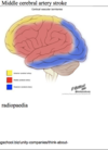
Symtoms would be focal (ie MCA distribution)
onset would be abrupt
pathologic changes would be ischemic
Include shrunken eosinophilic neurons acutely and later macrophages and gliosis
Neuron
most vulnerable cell
limited regeneration
Astrocyte
Major reactive cell of CNS (infarcts, neurodegenerative, infectious, ext)
proliferative
Oligodendrocyte
myelinated axonal processes
highly vulnerable (ie multiple sclerosis)
limited proliferation
Ependymal cell
lines ventricles
vulnerable
limited regeneration
Vascular disease cellular response
atherosclerosis, thrombosis, infarction/ eosinophilic neuronal necrosis
Red is dead = hypoxic, ischemic changes
Neoplastic cellular response
Unregulated proliferation of atypical cells due to tumor suppressor gene mutation
Neurodegenerative cellular response
Neuronal loss
gliosis
specific or non-specific inclusions (ie fibillary phosphotau positive inclusions; alzheimers or other neurodegenerative tauopathy)
Neurodegenerative cellular response 2
neuronal loss
subsequent loss of myelinated fibers in corticospinal tracts
ALS
Normal physiology
neuromelanin pigment
normal findings sumstantia migra/ locus ceruleus
Congenital inborn errors of metabolism cellular response
abnormal material within cytoplasm of neurons
neuronal storage diseases
Autoimmune cellular response
T cell inflammation
demyelination
macrophages
multiple sclerosis
Trauma cellular response
diffuse axonal injury shearing of axons/ vessels
axonal spheroids
hemorrhage
Toxic/ metabolic cellular response
alzheimers type II astrocytosis = hepatic encephalopathy
Infectious cellular response
Bacterial = neutrophils
Viral = microglial nodules/ lymphocytes
fungal = granulomas
rosenthal fibers
an be reactive (ie wall of a syrinx) or related to a pilocytic astrocytoma (pediatric tumor)
