Varus and Valgus deformities Flashcards
Infantile Blount's Adoslecent Blount's Genu algum
What are the causes of genu varum?
- Anchondroplasia
- Blount disease
- Trauma
- Infection
- Idopathic
- Osteogensis imperfecta
- Osteochondromas dysplasia
When is genu varum physiology?
- Normal in children less than 2 years
- Genu varum migrates to neutral at 14 months continue to genu valgum -knocked knees max at 3 years then back to neutral by 4 years
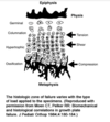
What is Blount’s disease?
- Is a progressive pathological genu varum centered at the tibia

Describe the 2 types?
-
Infantile- genu varum in children 0-3 years
- more common
- affects both lower limb extremities
- _ _Adolsecent- pathologcal genu varum in children >10 years
- usually unilateral
What is the aetiology of blount’s disease?
- Mutiltifactoral but related to mechanical overload in genetically susceptible individuals
- including excessive medial pressure produces an osteochondrosis of the medial prox tibial physis and epiphysis
- osteochondrosis -> physeal bar
What are the risk factors for blount’s disease?
- Overweight that are early walkers (<1 year)
what is the prognosis of blount’s disease?
- best outcomes with Early diagnosis and unloading of the medial joint with bracing or tibial osteotomy
What is the classification of Blount’s disease ?
- Lagenskiold
- Progress thru from I to 4 with increasing medial metaphyseal beaking and slope
- V and VI have epiphyseal - metaphyseal bony bridge- congenital bar across physis
- provides prognostic guidelines
What is often associated with Blount’s disease ?
- Internal tibial torsion, often bilateral
What is seen on examination of a child with Blount’s disease?
- Bilateral genu varum
- associated with internal Tibial torsion
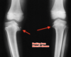
what is seen on X-ray?
-
Metaphyseal Beaking
- not seen in physiological bowing which is symmetrical flaring
- Asymmetric bowing
- progressive deformity
- varus focused at proximal tibia
- lateral thrust during gait

What angle is measured?
- Metaphyseal- diaphyseal angle of DRENNAN
- Angle between the line connecting metaphyseal beaks and line perpendicular to the longitudinal axis of the tibia
- >16 degrees is abnormal and 95% chance of PROGRESSION
- o 95 % of natural resolution of bowing

What is the tx of blount’s ?
Non operative
-
Brace tx with Knee ankle foot orthosis
- stage 1-2 in children <3yrs
- Metaphyseal-physeal angle 9-16o
- bracing must continue for 2 years for resolution of bony changes
- Outcomes
- good outcome with unilateral
- poor results with obesity and bilateral disease
Operative
-
proximal tibia/fibular valgus osteotomy
- stage 1-2 in children >3 years
- Stage 3-6 in children <3 years
- failure of bracing fx after 12 months
- rik of reocurrance less if preformed before 4 years
What are the goals of surgery?
- Overcorrect to 10-15o valgus as medial physeal growth abnormalities persist!!
- Distal segment is fixed in valgus, ext rotation and lateral translation
- Staples and screws increases forces across the physis which slows longitudinal growth- Heuter-Volkman principle= increasing compression across a growth plate leads to decreasing growth and increasing tension stimulates growth.
- resect physeal bar consider hemiepiphysiodesis of bar >50%
- medial tibial evaluation required at time of osteotomy
What is the technique of surgery?
staples and plates function by increasing compression forces across the physis which slows longitudinal plate= Heuter-voltman principles temporary lateral phsyeal growth arrest with plates/ staples include resection of bar (epiphysiololysis)
What do you see on examination of adolscent blount’s
- Genu varus bowing
- usually unilateral
- limb length discrepancy secondaty to deformity
- mild- moderate laxity of MCL

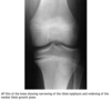
What is seen on xrays in adolescent blount’s?
- Metaphyseal beaking less common in adolescent cf infantile blount’s
- narrowing of tibial epiphysis
- widening of medial tibial growth plate
- occasional widening of the lateral distal femoral physis
What is the tx of adolescent blount’s disease?
Non operative
-
Observations
- mild
- poor outcome usually progresses and causes medial joi
- nt pain and altered kinmetics
- early onset arthritis typical
Operative
-
Lateral tibial and fibular epiphysiodesis
- mild to moderate with regrowth remaining
- up to 25% may need formal tibial osteotomy
-
Proximal tibial/fibular osteotomy
- more severe cases in skeletal maturity
- Distal femoral osteotomy or epiphysiodesis

Describe the surgical technique of a lateral tibia & fibular epiphysiodesis?
-
transient hemiepiphysiodesis
- tether physis with 8 plate or staples
- pros
- simple
- allows for gradual correction in children with adequate growth remaining
- implants may be removed
- dis
- requires significant growth remaining
- close observation required as growth plate may stop growing or rebound period of acceleration growth
-
permanent epiphysiodesis
- obliteration of physis thru lateral incision
- pros
- limited surgery
- overcorrection is uncommon
- doesn’t limit ability to do corrective osteotomy later
- disa
- can’t correct rotation
- up to 25% may require formal corrective osteotomy
What is the surgical technique of tibial & fibular ostoeotomy in adolscent blount’s?
-
High tibial osteotomy with rigid internal fixation
- ovecorrection is not indiciated cf infantile- just restore to neutral axis
- variety of techniques- open wedge/closing wedge then held staples
- limited weight bearing with crutches 6-8 wks
- pro
- immediate correction
- disc
- potential for compartment syndrome consider prophylaxtic fasciotomies
- quick lenthening of leg- neurological injury
Describe the surgical technique for ostetomy and slow correction in adolescent blount’s?
-
Tibial osteotomy and slow correction
- do osteotomy then attach frame- taylor spacial or ilizarov
- usually 12-18 week of tx required
- pro
- gradual correction reduced neurological compromise and risk of compartment syndrome
- allows for correction in all planes
- Dis
- pin site infection
- bulk of construct
- duration of tx
What are the causes of genu valgum in children?
It is important to distinguish normal physiological cf pathology
Bilateral
- Normal physiology
- Renal osetodystrophy ( renal Rickets)
-
Skeletal dysplasia
- Morquio syndrome
- spondyloeiphyseal dysplasia
- chondroctodermal dysplasia
Unilateral
- Physeal injury - trauma, infection or vascular insult
- proximal metaphseal tibial fx
-
benign tumours
- fibrous dysplasia
- Olliers disease
- Osteochondromas

Describe the normal physiology of genu valgum?
- Between 3-4 years = 20o genu valgum
- rarely worsens after 7 yrs
- after 7 yrs shouldn’t be >12o
- after 7 intermalleolar distance <8cm
What is the tx of genu valgum?
Non operative
-
Observation
- first line of tx
- genu valgum <15o in child <6 yrs
- Bracing
- rarely used
Operative
-
Hemiepephysiodesis or physeal tethering of bilateral medial femoral gorwth plates ( staples, screws or plate/screws, tension 8 plate)
- for >15-20o valgus in pt <10 years
- for femoral head to centre of ankle falls laterally in child >10 years
-
Distal femoral varus osteotomy- medial closing wedge
- insufficient growth for hemiepiphysiodesis
- distal femoral angle <88o ( normal 88o)
- complx
- peroneal nerve injury
- gradually correct the deformity
- utilizing closing wedge technique

