Elbow trauma Flashcards
Supracondylar fx Lateral epicondyle fx
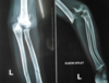
1
Q
What is this?

A
- An type 3 Garland supracondylar fx
- fall onto outstretched hand
- consisis of more than 1/2 of all paediatric elbow fx
- Extension type more common 95-98%
2
Q
what are the assoc injuires with supracondylar fx?
A
-
Anterior interosseous N neurapraxia ( branch median n)
- most common nerve palsy seen w SC fx
- weakness FDP index, FPL thumb- can’t make ok sign, weak FDP middle & pronator quadratus
-
Radial N palsy
- 2nd common neurapraxia
-
Ulna N palsy
- seen w flexion type SC- hand instrinsic weakness
- nearly all types of neurapraxia assoc with SC resolve spontaneously, so further dx studies not required
-
Vascular injury 1%
- Rich collateral circulation can maintain despite vascular injury
3
Q
Describe the age of ossification of the elbow epiphysis?
A
- Capitellum 1yr
- Radial head 4 yrs
- Internal (medial) epicondyle 6yrs
- Trochlear 8 yrs
- Olecranon 10 years
- Lateral Epicondyle 12 yr
** CRITOL**

4
Q
Describe the classification of elbow fx?
A
- Gartland
- Extension or Flexion type
-
Type 1
- non displaced
-
Type 2
- displaced, posterior cortex intact- see pic
-
Type 3
- completely displaced- open reduction & PP
-
Type 4
- complete periosteal disruption with instability in flexion/extension

5
Q
what are the signs of supracondylar fx?
A
-
AIN neurapraxia
- unable to flex IPJ of thumb and DIPJ of index finger - OK sign
-
Radial n palsy
- inability to extend wrist or digits
-
Vascular status
- at presentation 5-17%
- defined as cold, pale & pulseless hand
- a warm pink pulseless hand does not qualify as vascular insufficiency
- tx with immediate reduction and pinningi Theatre. attempted closed reduction in ER first

6
Q
What are you looking for on radiographs with a SC fx?
A
- Anterior humeral line should intersect the middle third of the capitellum
- Capetilum moves posteriorly to this reference line in an Extension type SC fx
- Baumann angle
- created by a line perpendicular to humeral shaft adn a line along the lateral condylar physis as viewed on ap image
- normal 85-89 degrees cf contralteral side
- deviation of 5 degrees= coronal plane deformity & should not be accepted

7
Q
What is the tx of Sc fx?
A
Non operative
- type 1
- type 2- ant humeral line insects capitellum, minimal swelling, no medial comminution
- Posterior moulded splint then long arm cast at 90o or less
- typically for 3 weeks
Surgery
-
Closed reduction & percutanous pinning
- _ _most supracondylar fx
- Lateral fragments- supinatn w hyperflexion
- medial fragments- pronation w hyperflexion
- 2 lateral pins usually sufficient
- confirm stability
-
crossed pins
- strongest to torsional stress in expt studies
- > risk of ulnar n injury 3-8%
- remove pins 3 weeks
-
Open reduction and percutanous pinning
- reduction not obtained closed
- anterior approach
8
Q
Describe the tx of pulseless, cool hand or floating elbow from Sc fx?
A
-
Immediate closed reduction and percutaneous pinning
- check vascular status after reduction
- explore if pulse if lost after reduction or if pulseless, plae hand persists after reduction
- anteriography is typically not indicated
9
Q
Describe the complications of Sc fx?
A
-
Pin migration
- most common complication 2%
-
Infection
- 1%
- superfically, oral antibiotics
-
Cubitus Valgus
- caused by fx malunion
- -> tardy ulnar nerve palsy
-
Cubitus varus
- gunstock deformity
- caused by fx malunion
- -> fx, cosmetic issue w little functional limitation
-
Recurvatum
- __common w non op Type 2/3 fx
-
Nerve palsy
- usually resolves
- Vascular injury
-
Volkmann ischaemic contracture
- rare
- normally result of brachial artery compression and tx utilzing elbow hyperflexion and casting then true arterial injury
- increase in forearm compartment pressures and loss of radial pulse w elbow flexed greater than 90o
- Post op stiffness

10
Q
What is a floating elbow?
A
- ipislateral supracondylar humerus and a forearm fracture
- must be operated on emergency to reduce risk of compartment syndrome
- wrist pinned first to allow fulcrum for reduction of supracondylar fx
11
Q
Which is the last apophysis to fuse in the elbow?
A
- Medial epicondyle
- aged 16-19 years
12
Q
What is this?

A
- A Milch 2 lateral condylar fx
- 2nd most common pediatric elbow fx
- 6 yrs most common
- prognosis:
- outcomes have been historically worse than Supracondylar fx
- articular nature, missed dx, higher risk of malunion/nonunion
13
Q
What is the classification of lateral condyle fx?
A
- Milch
-
Type 1
- fracture line is lateral to trochlear groove
- considered SH IV
-
Type 2
- FX line INTO trochlear Groove
- Consider SH 2 fx

14
Q
What are the signs and symptoms of lateral condyle fx?
A
- elbow pain
- swelling and tenderness limited to lateral side
15
Q
What images are useful in lat condyle fx?
A
- Xrays
- Ap
- lateral
- internal oblique view= shows max displacement & fx pattern
16
Q
What is the for lateral condyle fx?
A
Non operative
- if <2mm displacement
- cartilagenous hinge intact
- weekly FU
Surgery
-
Closed reduction & percutaneous pinning
- w no evidence of intra-articular incongruity
- divergent pin placed most stable
-
Open reduction and percutanous pinning
- if >2mm displacement
- any joint incongruity
- direct lateral approach
- avoid dissesction of posterior aspect of lateral condyle ( source of vascularisation)
17
Q
What are the complications from lateral condyle fx?
A
-
AVN
- posterior dissection -> lat condyle necrosis
- may also occur at trochlea
-
Non union/malunion
- caused a delay in diagnosis and improper tx
- -> cubital valgus and tardy ulna n palsy
- if symptomatic screw fix & bone graft non union
-
Lateral overgrowth/prominence spurring
- in up to 50% cases reagrdless of tx
- lateral periosteal alignment will prevent this from occuring
- prsence of spurring is correlatd with greater intial fx displacement
- Growth arrest w /wout angular deformity
- unsatisfactory appearance of surgical scar

