Developmental hip dysplasia Flashcards
what is developmental dysplasia of the hip?
- A disorder of abnormal development resulting in dysplasia and possible subluxation or dislocation of the hip secondary to capsular laxity and mechanical factors
What does DDH encompass?
- A spectrum of disease that includes
- DYSPLASIA
- SUBLUXATION
- DISLOCATION
- TERATOLOGIC HIP- identified in utero- has pseudoacrtabulum, assoc with arthrogryposis, myelomeningocele
- LATE ADOLESCENT
What is the incidence?
- Hip dysplasia 25%
- scotland 4 per 1000 live births
- bilateral in 20%
- most common left hip in females
What are the risk factors?
- First born
- female 6:1 males
- breech
- oligohydraminos
- FHx
- assoc with other packaging deformities
- congential muscular torticolis 20%
- Metatarsus adductus 10%
- congenital knee dislocation
What is the pathology meant to be caused by?
- INITAL instability thought to be due to maternal and fetal laxity
- Genetic laxity
- Intrauterine and postnatal malpositioning
- initial laxity-> dysplasia -> gradual dislocation
Where is the normal acetabular deficiency ?
- Anterior or anterolateral
- In cerebral palsy= posteriosuperior
What is the classification?
- Put into a spectrum of diseases
-
DISLOCATED
- Ortani positive- reduce hip
-
DISLOCATABLE
- Barlow positive
-
SUBLUXABLE
- Barlow positive
-
DISLOCATED
How do you diagnose it?
Physical exam <3 months
- ORTOLANI- hip is OUT AND TRY AND REDUCE IT- abduction manoeuvre
- BARLOW- hip is IN and try push back to dislocate it
-
GALEAZZI- to identify any LLD due to unilate dislocated hip with hip and knees flexed
- 90 degrees femur short on dislocated hip
In a child older than 3 months what would your clinical findings be in a child with DDH?
-
Limited HIP ABDUCTION
- most sensitive test when contractures start
- LLD Unequal skin folds- only in unilateral
- >1 yr when walking
- pelvic obliquity
- lumbar lordosis
- trendlenberg gait- abductor insufficiency
- toe walking - compensate for shortening of affected side

What age can you use radiographs? why?
- From 4-6 months
- as this is when the FEMORAL HEAD begins to OSSIFY
-
Tear drop seen at 18 months
- this is quadrilateral space & cotyloid fossa
- if seen within 6 months of reduction of hip strongly good outcome

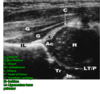
What lines on X-ray help you identify hip dislocation in DDH?
-
Hilgenreiner’s line= line thru r + l triradiate cartilage
- Femoral head ossification should N be INFERIOR
-
Perkins line= line perpendicular to hilgenreiner’s thru a point at a LATERAL margin of the ACETABULUM
- Femoral head ossification should N be medial to this line
-
SHENTON’S line- arc along inferior border of femoral neck and sup margin of obturator foramen
- arc should be N continuous

What lines on X-ray help you identify hip dysplasia in DDH?
-
Acetabular Index- angle formed by a line drawn from point on the lateral triradiate cartilage to point on lateral margin of acetabulum and hilgenreiner’s line
- should be < 25 degrees in pt>6/12
-
CENTRE EDGE ANGLE of WILBERG
- angle formed by a vertical line from the centre of the femoral head and a line from the centre of the femoral head to the lateral edge of the acetabulum
- N is >20 degrees only reliable after 5 yrs

When is USS recommended?
- At 4-6 weeks in baby with
- risk factors
- positive physical findings
- utilized follow Pavlik tx for equivocal exams
What is the use of USS in DDH?
- it is DYNAMIC USS useful before femoral head ossifies at 4-6 months to identify the ALPHA ANGLE
- Alpha angle created by lines along the bony acetabulum and ilium
- normal >60degrees
- BETA ANGLE - angle created by lines along labrum and ilium, normal <55 degrees
- USED for assessing reduction of hip whilst in PAVLIK HARNESS- avoids unreduced hips eroding acetabulum
- Not cost effective for routine screening

What is the goal of tx?
- Based on Achieving and Maintaining early concentric REDUCTION in order to prevent FUTURE degenerative joint disease
- Tx is based on child’s age
When is arthrogram useful in DDH?
- Used to confirm reduction after closed reduction under anesthesia
- Concentric reduction must be obtained with <5mm of contrast pooling medial to femoral head and the limbus must not be interposed
- Helps to identify blocks to reduction
- Inverted Labrum
- labrum reduces depth of acetabulum by 20-50% adn contributed to growth of acetabular rim
- older child may be inverted and block cocentric reduction of the hip
- Inverted limbus- rosethorn sign
- a pathologic response of acetabulum to abnormal pressure caused by superior migration of the head
- consists of fibrous tissue
- Pulvinar
- ligamentum teres
- hip capsule constricted by iliopsoas tendon causing hour- glass deformity pf capsule

When is Ct useful in DDH?
- Ct study of choice to evaulate reduction after closed reduction and spica casting
- MRI doesn’t play a significant role in primary diagnosis
What is tx birth to 6 months ?
- HIP MUST BE REDUCIBLE
- Pavlik harness- abduction splinting
- check at 3 weeks USS to confirm femoral head reduction
- Requires normal muscle function CI in pt with spina bifidia
- Outcomes
- success rate of 90%
- abandon brace if not successful at 3-4 weeks

What happens if no reduction on uss at 3 weeks in a child 0-6 months?
- Discontinue Pavlik harness to prevent pavlik disease
- Erosion of pelvis superior to acetabulum -> difficulty with closed reduction
- Consider closed reduction under GA with on table arthrogram as femoral head is not ossified. then spica cast post op MRI to confirm concentric reduction
What is tx 6-18 months with a reducible hip ?
-
Closed reduction under GA + arthrogram and spica casting
- arthrogram allows to see femoral head/any obstuctions to reduction
- Possible adductor tenotomy
- Spica cast
- Post op MRI to confirm concentric reduction
What happens if this closed reduction fails or 6-18month old unreducable hip? Why does closed reduction fail?
- Then open reduction and spica cast
- Due to
- INVERTED limbus
- Pulvinar
- HYPERTROPHIED Ligamentum teres
- ILIOPSOAS CONTRACTURE
- Capsular constriction
What is tx >2 years with residual hip dysplasia ?
-
Open reduction and Femoral oseotomy
- Osteotomy to correct femoral anteversion and coxa valga
- Also prevent AVN
- aim is to congruently reduce the femoral head with GROM
- May add in pelvic osteotomy often > 4yrs and if severe dysplasia accompanied by significant radiographic changes on acetabular side ie. Acetabular index >25 degrees
What approach is used to open reduce the hip?
- Anterior- smith peterson
- As less risk of injury to medial femoral circumflex vessel
Could a medial approach be used?
- Yes
- Advantages can be used in child <12 months
- directly addresses block to reduction.
- less blood loss but disadvantages- unable to preform capsulorrhapy and high associated risk of AVN
What is the position of the childs hips in the pavlik harness?
- Within safe zone
- Flexion 90-100 degrees- control anterior straps
- Abduction 50 degrees- control posterior straps
What can positions outwith the safe zone cause?
- Abduction >60
- Causes
- **AVN **
- impingment of posteriorsuperior retinacular branch of middle femoral circumflex artery
-
transient femoral n palsy
- due to hyperflexion
- **AVN **
How long is the harness worn for?
- 23 hours a day for at least 6 weeks
- uss at 3 weeks to confirm reduced
- wean out of harness over 6-8 weeks after hip has stabilised
- confirm hip position with uss every 4-6 weeks
What position in the hip placed in the spica cast?
How long do they remain in it?
How frequently are they changed?
- flexion 100 degrees
- abduction 45 degrees
- Remain in spica for 3 months
- Changed every every 6 weeks
What pelvic osteotomy would you use in a young child?
- Salter Open triradiate cartilage
- single osteotomy cut above acetabulum thru ILIUM to sciatic notch acetabulum
- hinges thru pubic symphysis
- can provide 20-25 degrees lateral and 10-15 anterior coverage may lengthen up to 1cm

What pelvic osteotomy would you use in an older child?
- Triple STEELE
- Salter osteotomy plus inferior and superior rami cut
- acetabular reorientation proceedure
- triradiate cartilage must be open
- Reorientation proceedure

What pelvic osteotomy would you use in a child > 8 years with a closed triradiate cartilage?
- Ganz or A Shelf salvage proeceedure
- Ganz- multiple ostetomies in the pelvis, ilium and ischium nr the acetbulum- improves 3d correction but technically the most challenging .
- SHELF- when you add bone to the lateral WB surface of the acetabulum by extra-articular buttress of bone over sublimed femoral head. it depends on the fibrocartilage metaplasia for successful results

What the general complications of DDH tx?
-
Osteonecrosis
- All forms of tx
- excessive/ forceful abduction
- previous failed closed tx
- repeat surgery
- seen on xray- failure of growth of ossificaiton nucleus 1yr after reduction
- broadening of femoral neck
- increased density & fragmentation of ossified head
-
Recurrence
- Approx 10% with appropriate tx
- radiological follow up until skeletal maturity
- Delayed diagnosis
What if a child presents late with bilateral dislocations if aged 6 or older?
- Pt typically functions better if hips NOT reduced
What if a child presents late with unilateral dislocation if aged 8 or older?
What can be done about their LLD?
- Better outcomes without surgical tx if patient aged 8 or more
- Epiphysiodesis
