The Respiratory System Flashcards

1
Q

RESPIRATORY TRACT

Upper

  • Humidify & warm air
  • Defence – mucus and cilia
  • Sensory
  • Speech

Lower

  • GAS EXCHANGE
  • Defence
  • Metabolic
A
2
Q

WHAT IS THE FUNCTION OF THE LARYNX?

A

To generate different sounds

3
Q

WHAT PARTS OF THE LUNGS ARE KNOWN AS THE ‘CONDUCTING ZONES’?

A

The tubes (e.g. trachea and primary bronchus)

4
Q

LOWER RESPIRATORY TRACT

A
5
Q

WHAT IS THE PARENCHYMA?

A

The part of the lungs where gas exchange occurs

6
Q

LUNG BRANCHES

A
7
Q

AIRWAY RESISTANCE

  • Proportional to 1/radius4
  • But conduction summative

–huge numbers of respiratory bronchioles

–small airways make a small contribution to total Resistance

•Conducting zone bronchioles largest influence

Inversely proportional to conductance; higher the resistance, the less efficient conduction.

A
8
Q

AIRWAY RESISTANCE IS INCREASED IN MANY DISEASES

  • Smooth muscle contraction
  • Obstruction with mucus
  • Collapse
  • Structural changes occur in the conducting airways
  • Smooth muscle growth
  • Fibrosis
A
9
Q

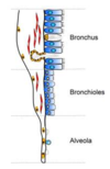
LUNG EPITHELIUM

  • Lining cells in airway
  • Specialisation changes down the airway

Bronchi

•ciliated, goblet, glandular

Bronchioles

•ciliated, non-ciliated, goblet, club cells

Alveoli

•Squamous, cuboidal

Ciliated= waft

Goblet cells secrete mucus (as do glandular cells)

Club cells (secretory cells)

Highly specialised cells in the alveoli (specialised for gas exchange).
A
10
Q

AIRWAY STRUCTURE

A
11
Q

AIRWAY INNERVATION

Sensory (afferent) nerves FROM the airway epithelium and smooth muscles signal to the brain.

Autonomic fibres TO glandular epithelium and smooth muscle from CNS

  • Parasympathetic branches of the vagus nerve (vagus nerve is the main nerve supplying the lungs).
  • Excitatory (e) and inhibitory (i)
  • Very little sympathetic innervation (most nerves are parasympathetic).
  • ß-adrenoceptors on airway smooth muscle are stimulated by circulating adrenaline.
A
12
Q

THE RESPIRATORY UNIT

•Massive network of airways and air sacs (around 500 million) to maximise surface area (50-100 m2).

Pneumocytes are very thin, flat cells to allow maximum _ exchange.

Also surrounded by dense _ network= gas exchange.

A

Capillary

Gas

13
Q

ALVEOLAR EPITHELIUM CELLS (PNEUMOCYTES)

•Type I (Gas exchange cells)

–Very large surface area

  • cover 95% of alveolus
  • 10% of cells

–squamous

–gas exchange

•Type II cells (secrete surfactant, they also replace TI cells when the TI cells die)

–cuboidal

•majority of cells

–secretory

•surfactant

–precursors for type I

A
14
Q

WHAT DO TYPE II CELLS SECRETE?

A

Surfactant

15
Q

SURFACTANT

•Produced by type II alveolar epithelial cells

–Lipid (90%) – mostly phospholipid

–Proteins – SP-A, -B, -C, -D

•Reduces surface tension

–prevents alveolar collapse (atelectasis)

  • Innate immunity function
  • Instilled into babies with Infant Respiratory Distress Syndrome

–Beractant (bovine)

–Pumactant (synthetic, lipid only)

A
16
Q

LUNGS AT BIRTH

  • Lung epithelium develops in last trimester of pregnancy.
  • Maturation stimulated by _ in premature babies
  • Lung is fluid filled in foetus - must rapidly empty

–Surge in corticosteroids and catecholamines at birth

–Activation of absorptive channels

•Epithelial Sodium Channels (ENaC)

–Pressure changes (squeeze through birth canal)

•Caesarian section – reduced drive for fluid absorption

A

Corticosteroids

17
Q

BLOOD SUPPLY TO THE LUNGS

•Pulmonary circulation

–Artery flows directly from the right ventricle

  • LOW oxygen
  • HIGH flow
  • _ pressure

–Capillaries pass around the alveoli

–Pulmonary _ returns oxygenated blood to left heart

•Bronchial circulation

–From aorta (left ventricle)

–HIGH pressure

–Supplies oxygen and nutrients the conducting airways

–Not involved systemic respiration processes

–Only 2% of cardiac outflow

A

Low

Vein

18
Q

VENTILATION IS AUTOMATIC AND CONTROLLED BY THE CNS.

TRUE OR FALSE?

A

TRUE

19
Q

NEURONAL CONTROL OF BREATHING

A
20
Q
A
21
Q

MUSCLE INNERVATION

Somatic motor nerves

Innervate skeletal muscles in the thorax

  • Phrenic
  • innervates the diaphragm
  • irritation => hiccough
  • Intercostal

Autonomic nerves

Bronchial

•supply smooth muscle & secretory cells

–branch from vagus

–reflex bronchospasm and mucus secretion

–important in asthma

Sensory output

A
22
Q

RESPIRATORY MUSCLES

Intercostals are between the _.

External intercostals contract to lift ribs _.

Internal intercostals contract to pull ribs down again and to force air out.

A

Ribs

Upwards

23
Q

SENSORY AFFERENT PATHWAYS FROM THE LUNG (MYELINATED)

•Send impulses via vagus to medullary centres

Slowly adapting

–Stimulated by stretch receptors in airway smooth muscle

–Elicit reflexes

  • Shortened inspiration
  • Hering-Breuer reflex

–Promotion of expiration following steady inflation

–Prevents over-inflation of lung

Rapidly adapting stretch receptors

–Stimulated by sudden, sustained inflation

–Also by ‘irritant receptors’ among epithelium

–Elicit reflexes:

  • Cough
  • Bronchoconstriction
  • Mucus secretion
A
24
Q

WHAT STIMULATES RAPIDLY ADAPTING STRETCH RECEPTORS?

A

Sudden, sustained inflation of the lungs

25
Q

SENSORY AFFERENT PATHWAYS (NON-MYELINATED FIBRES)

  • Pulmonary and bronchial C fibres
  • Located close to blood vessels (J receptors)
  • Exogenous stimuli

–Noxious agents in air

•Endogenous stimuli

–inflammatory agents generated by the body

•Reflex bronchoconstriction & mucus secretion

A
26
Q

WHAT DO J RECEPTORS CAUSE?

A

Cause changes in blood flow in specialised regions

27
Q

COUGH REFLEX

•Stimulation of irritant receptors

–Sensory nerves conveys signal to medulla

  • Motor nerves signal to _ muscles
  • Glottis closes
  • Abdominal and _ intercostal muscles contract rapidly
  • Intrapulmonary pressure rises
  • Glottis opens
A

Skeletal

Internal

28
Q

PLEURAL MEMBRANES

  • Double membrane surrounding the lungs
  • Left & right pleura anatomically distinct

–Collapsed lung (pneumothorax) usually only affects one lung

•Normally very small amounts of pleural fluid between membranes (few ml)

–Serous fluid

•Lubricant

A
29
Q

VENTILATION

  • Fluid in the pleural sac helps the lung wall “stick” to the inside of the thorax.
  • Contraction of the _ and the external intercostal muscles.

–Increased lung _.

–Internal pressure falls (Boyles Law)

–Air drawn into lungs

•Exhalation is passive at rest

–Elastic recoil

A

Diaphragm

Volume

30
Q

IN TERMS OF VENTILATION, WHAT HAPPENS WHEN YOUR DIAPHRAGM AND EXTERNAL INTERCOSTAL MUSCLES CONTRACT?

A
  1. Lung volume increases
  2. Internal pressure falls
  3. Air is drawn into the lungs
31
Q

PULMONARY COMPLIANCE

A
32
Q

METABOLIC FUNCTION OF THE LUNG

•Club cells _ inhaled substances

–cyt p450

•Vascular cells inactivates some circulating hormones

–prostaglandins

•Vascular cells activate angiotensin I

–Biologically inert Ang I converted to pressor Ang II

(Angiotensin Converting Enzyme)

•Fibrinolytic function

A

Detoxify

33
Q

WHAT DO CLUB CELLS DO IN THE LUNGS?

A

Detoxify inhaled substances

34
Q

NEURAL AND CHEMICAL CONTROL OF RESPIRATION

Neural control

Central rhythm generator in medulla

  • Receptors in respiratory tract causing sneezing, coughing and hyperpnoea
  • Nociceptors

Chemical control

•Central and _ chemoreceptors

A

Peripheral

35
Q

NEURONAL CONTROL OF BREATHING

Medulla

–Ventral and Dorsal Respiratory Group:

  • Discharge rhythmically
  • efferent neurons to motor nerves
  • receives afferent input from periphery and pons
  • Little activity in expiratory centre at rest

Pons

–Apneustic centre:

  • Prolongs medullary centre firing
  • Hence depth of breathing increased

–Pneumotaxic centre:

  • Inhibits apneustic centre
  • Controls rate of breathing
A
36
Q

WHAT DOES THE APNEUSTIC CENTRE IN THE BRAIN DO?

A

Prolongs medullary centre firing= increases depth of breathing

37
Q

WHAT DOES THE PNEUMOTAXIC CENTRE OF THE BRAIN DO?

A

Inhibits apneustic centre

Controls rate of breathing

38
Q

RHYTHMIC DISCHARGE OF PRE-BÖTZINGER COMPLEX

  • Region of the ventral respiratory group in the _.
  • Spontaneous rhythmic discharge.
  • Stimulates rhythmic discharge of motor nerves.
  • Results in contraction of _.
A

Medulla

Diaphragm

39
Q

WHAT IS ONDINE’S CURSE?

A

Loss of automatic control of ventilation

40
Q

VOLUNTARY CONTROL OF BREATHING

Via the Cerebral Cortex.

  • Sends signals direct to respiratory motor neurones
  • Sensitive to:

–Temperature

–Emotion

A
41
Q

LUNG TRANSPLANT

  • Motor innervation is to skeletal muscles, so ventilation maintained
  • Preservation of cough from tracheal stimulation
  • Loss of cough stimulation from lower airway
  • Loss of Hering-Breuer reflex
A
42
Q

WHAT TWO TYPES OF RECEPTORS CONTROL RESPIRATION?

A

Central Chemoreceptors (respond to [H+] and also the concentration of CO2)

Peripheral Chemoreceptors- Carotid and Aortic bodies (Primary signal is O2 but there is also some input from [H+].

43
Q

WHAT IS THE NORMAL AIR CONCENTRATION OF CO2?

A

0.04% (0.3 mm Hg)

44
Q

WHAT IS THE NORMAL ATMOSPHERIC CONCENTRATION OF OXYGEN IN THE AIR?

A

21% (160mm Hg)

45
Q

WHAT DOES PaO2 AND ALSO PaCO2 STAND FOR?

A

Partial pressure of arterial oxygen (normally about 80-100mm Hg)

Partial pressure of arterial CO2 (normally about 40mm Hg)

46
Q

WHAT DOES PAO2 STAND FOR?

A

Concentration of alveolar oxygen

47
Q

CENTRAL CONTROL OF RESPIRATION

Carbon Dioxide Stimulates the Chemosensitive Area

Although carbon dioxide has little direct effect in stimulating the neurons in the chemosensitive area, it does have a potent indirect effect. It does this by reacting with the _ of the tissues to form carbonic acid, which dissociates into _ and bicarbonate ions; the hydrogen ions then have a potent direct stimulatory effect on respiration.

Why does blood carbon dioxide have a more potent effect in stimulating the chemosensitive neurons than do blood hydrogen ions?

The blood-brain barrier is not very permeable to hydrogen ions, but carbon dioxide passes through this barrier almost as if the barrier did not exist. Consequently, whenever the blood PCO2 increases, so does the PCO2 of both the interstitial fluid of the _ and the cerebrospinal fluid. In both these fluids, the carbon dioxide immediately reacts with the water to form new hydrogen ions. Thus, paradoxically, more hydrogen ions are released into the respiratory chemosensitive sensory area of the medulla when the blood carbon dioxide concentration increases than when the blood hydrogen ion concentration increases. For this reason, respiratory center activity is increased very strongly by changes in blood carbon dioxide levels.

(PIC SHOWS WHAT OCCURS IN THE CSF)

A

Water

Hydrogen

Medulla

48
Q

HOW DOES CARBON DIOXIDE (INDIRECTLY) STIMULATE THE NEURONS IN THE CHEMOSENSITIVE AREA?

A

. It does this by reacting with the water of the tissues to form carbonic acid, which dissociates into hydrogen and bicarbonate ions; the hydrogen ions then have a potent direct stimulatory effect on respiration

49
Q

THE BLOOD-BRAIN BARRIER IS VERY PERMEABLE TO HYDROGEN IONS BUT NOT TO CARBON DIOXIDE.

TRUE OR FALSE?

A

FALSE

It isn’t very permeable to hydrogen ions but carbon dioxide can pass through it a though it almost doesn’t exist.

50
Q

WHAT DOES CSF STAND FOR?

A

Cerebrospinal fluid

51
Q

HENDERSON-HASSELBACH EQUATION

(TURN OVER TOO)

A
52
Q

REGULATION OF CO2 DURING SUSTAINABLE EXERCISE

•Ventilation increases prior to rise in blood CO2

The presumed reason that the ventilation forges ahead of the buildup of blood carbon dioxide is that the brain provides an “_” stimulation of respiration at the onset of exercise, causing extra alveolar ventilation even before it is necessary. However, after about 30 to 40 seconds, the amount of carbon dioxide released into the blood from the active muscles approximately matches the increased rate of _, and the arterial PCO2 returns essentially to normal even as the exercise continues.

  • Arterial PCO2 levels fall
  • As exercise proceeds, more CO2 generated
  • CO2 passes from muscles into venous _
  • Efficiently removed in lung
  • When exercise ceases, ventilation falls rapidly
A

Anticipatory

Ventilation

Blood

53
Q

PERIPHERAL CONTROL RESPIRATION

Fall in O2 stimulates glomus cells in carotid and aortic bodies.

  • Contain O2 sensitive K+ channels and dopamine
  • O2 fall
  • O2 sensitive K+ channels CLOSE
  • Depolarisation
  • DA release
  • Stimulates afferent fibres
  • Signals to medulla
A
54
Q

REGULATION OF AFFERENT FIBRE ACTIVITY FROM THE CAROTID BODY BY O2

A
55
Q

RESPIRATORY STIMULANTS

Doxapram

Closes K+ channels on glomus cell (carotid body)

•Glomus cell depolarises

  • sends afferent signals to medullary respiratory centre

Central action at high dose

Use in respiratory failure (rarely)

Caffeine (& other xanthines)

–Non-specific CNS stimulation, including respiratory centre

–Bronchodilator (via phosphodiesterase inhibition?)

–Used in sleep apnoea and premature babies.

Acetazolamide

Carbonic anhydrase inhibitor. Stimulates respiration by creating mild metabolic acidosis via decreased renal reabsorption of bicarbonate and hence reduced acid buffering.

Carotid bodies respond to decreased pH (although fall O2 is primary drive)

A
56
Q

WHAT ARE THREE EXAMPLES OF RESPIRATORY STIMULANTS?

A

Doxapram

Caffeine

Acetazolamide

57
Q

ALCOHOL IS A STIMULANT.

TRUE OR FALSE?

A

FALSE

It is a depressant

58
Q

RESPIRATORY DEPRESSANTS

A
59
Q

WHY DO WE BREATHE?

•O2 needed for aerobic respiration

–Krebs’ cycle cannot proceed without regeneration of _.

–_ Transport Chain crucially dependent on O2

•ATP generated by respiration required for all active processes

–Active transport

  • Ionic gradients
  • Nutrient uptake

–Anabolic reactions

–Muscle contraction

–Phosphorylation of targets

–Precursor for cAMP

•O2 also needed to pay ‘oxygen debt’ after bursts of _ metabolism

A

Anaerobic

NAD+

Electron

60
Q

PARTIAL PRESSURE OF GAS

A
61
Q
A
62
Q

HOW MUCH O2 IS CONSUMED?

A
63
Q

BLOOD TRANSPORT

CO2 production measured in arterial blood

•Normal arterial PCO2 is about 37 mm Hg

so Alveolar oxygen pressure

PAO2 = O2 inspired – O2 consumed

= O2 inspired – (CO2 produced/RQ)

= 150 – (37/0.8) mmHg

= 104 mm Hg

A
64
Q

DIFFUSION OF OXYGEN INTO TISSUES

A
65
Q

HOW MANY OXYGEN MOLECULES CAN ONE HAEMOGLOBIN MOLECULE BIND?

A

4

66
Q

OXYGEN RELEASE IN TISSUES

  • As oxygenated arterial blood reaches capillaries, O2 released
  • Consumed by cells
  • Arterial PO2 100 mmHg
  • Venous PO2 40 mmHg
A
67
Q

HAEMOGLOBIN BINDS CO-OPERATIVELY.

TRUE OR FALSE?

A

TRUE

68
Q
A
69
Q

CO2 TRANSPORT

  • CO2 transported mainly (70%) as HCO3-
  • 7% as dissolved _
  • 23% as carbamino haemoglobin (HbCO2)
  • CO2 from tissues diffuses into plasma.
  • Most of this (80%) enters red blood cells and is converted to bicarbonate by carbonic _.
  • When CO2 production increased (exercise or disease)
  • Fraction of CO2 increases relative to HCO3-
  • Passage into CSF and detection by central _.
A

CO2

Anhydrase

Chemoreceptors

70
Q

WHAT IS MOST CO2 (70%) TRANSPORTED IN THE FORM OF?

A

HCO3-

71
Q

GAS PRESSURES IN ALVEOLI AND BLOOD

•PO2 same in alveolus and pulmonary vein, but much higher than in systemic veins (and hence the pulmonary artery)

•PCO2 higher in veins than alveolus and systemic arterial blood

A
72
Q

GAS PRESSURES IN ALVEOLI AND BLOOD

  • PO2 same in alveolus and pulmonary vein, but much higher than in systemic veins (and hence the pulmonary artery)
  • PCO2 higher in veins than alveolus and systemic arterial blood
A
73
Q

WHAT HAPPENS WHEN OXYGEN BINDS TO HAEMOGLOBIN?

A
  • Haemoglobin becomes more acidic
  • Less formation of Hb.CO2
  • Excess H+ bind bicarbonate to form carbonic acid
  • CO2 released
74
Q

VENTILATION-PERFUSION MATCHING

A
75
Q

WHAT IS HYPOXAEMIA?

A

Low oxygen levels

76
Q

WHAT IS HYPERCAPNIA?

A

Increased CO2 levels

77
Q

CAUSES OF HYPOXAEMIA

•Shunting

–Anatomical

–Unventilated alveoli

  • Ventilation-perfusion mismatch
  • Diffusion abnormalities

–thickened alveolar wall

•Hypoventilation

–Neuronal defect or muscle weakness

–Obstruction

–drug-induced

CAUSES OF HYPERCAPNIA

Increased dead space

-pulmonary embolus

Hypoventilation

A
78
Q

PHYSIOLOGICAL RESPONSE TO LOW INSPIRED OXYGEN

•Increased ventilation (immediate)

–Driven by peripheral chemosensors

–Not sustained

•Pulmonary vasoconstriction (rapid and sustained)

–Homeostatic mechanism designed to shunt blood away from poorly ventilated areas

–Not useful if the whole lung is hypoxic!

•Increased haematocrit (red cell fraction of blood > 50%)

–HIF (hypoxia inducible factors) activation

–HIF regulates oxygen-sensitive gene transcription

  • Erythropoetin production by kidneys
  • EPO stimulates bone marrow to make red blood cells
A
79
Q

WHAT ARE THE THREE PHYSIOLOGICAL RESPONSES TO LOW INSPIRED OXYGEN?

A

Increased ventilation

Pulmonary vasoconstriction

Increased haematocrit

80
Q

ALTITUDE SICKNESS

A